 Indietro
Anatomy & Physiology Study Guide: Sense Organs (Hearing & Equilibrium, Vision)
Guida di studio - Note intelligenti
 Appunti personalizzati basati sui tuoi materiali, ampliati con definizioni chiave, esempi e contesto.
Sense Organs
Sensory Reception
Sensory reception is the process by which specialized structures called sensory receptors detect environmental stimuli and convert them into neural signals. These receptors are classified based on the type and origin of the stimulus.
Thermoreceptors: Detect temperature changes.
Photoreceptors: Detect light (e.g., vision).
Nociceptors: Detect pain.
Chemoreceptors: Detect chemical changes (e.g., ions, pH, gas levels).
Mechanoreceptors: Detect physical changes (e.g., touch, pressure, stretch, vibration, tension).
Exteroceptors: Sense stimuli external to the body.
Interoceptors: Sense stimuli internal to the body.
Proprioceptors: Sense body position and movement.
Hearing & Equilibrium
The Ear: Anatomy
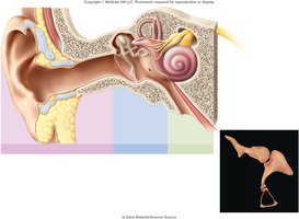
The ear is divided into three distinct regions: the outer ear, middle ear, and inner ear. Each region plays a specific role in the process of hearing and balance.
Outer Ear
The outer ear acts as a funnel to conduct sound vibrations to the eardrum. It consists of:
Auricle (Pinna): Fleshy projection supported by elastic cartilage.
Auditory Canal (External Auditory Meatus): Passage leading to the tympanic membrane.
Cerumen (Earwax): Produced by ceruminous and sebaceous glands; protects, waterproofs, and keeps the tympanic membrane pliable.
Middle Ear
The middle ear is located in the tympanic cavity of the temporal bone and conducts and amplifies sound. It contains:
Tympanic Membrane (Eardrum): Boundary between outer and middle ear; vibrates in response to sound.
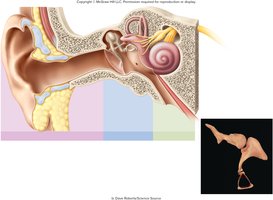
Auditory Ossicles: Three bones (malleus, incus, stapes) that transmit and amplify sound vibrations.
Auditory (Eustachian) Tube: Connects middle ear to nasopharynx; equalizes air pressure and drains the middle ear.
Inner Ear
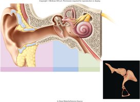
The inner ear is housed in the bony labyrinth of the temporal bone and functions in hearing and equilibrium. It consists of:
Bony Labyrinth: Passageways carved into the temporal bone.
Membranous Labyrinth: Soft tissue tubes lining the bony labyrinth.
Perilymph: Fluid between bony and membranous labyrinth (similar to CSF).
Endolymph: Fluid within membranous labyrinth (similar to ICF).
The Cochlea
The cochlea is a coiled, snail-shaped organ comprised of three fluid-filled chambers separated by membranes. It is essential for hearing.
Scala Vestibuli: Superior chamber starting at the oval window; filled with perilymph.
Scala Media (Cochlear Duct): Middle chamber filled with endolymph; contains the spiral organ.
Scala Tympani: Inferior chamber ending at the round window; filled with perilymph.
Vestibular Membrane: Separates scala vestibuli from scala media.
Basilar Membrane: Separates scala media from scala tympani.
Tectorial Membrane: Gelatinous layer on top of spiral organ.
Spiral Organ (Organ of Corti): Thick epithelial layer of hair cells with stereocilia; converts sound vibrations into nerve impulses.
Physiology of Hearing
Sound waves are funneled inward by the outer ear, causing vibration of the auditory ossicles in the middle ear. This movement leads to fluid motion in the cochlea, ultimately resulting in the opening of mechanically gated ion channels in hair cells.
Mechanically Gated Ion Channels: Vibration of the basilar membrane pushes stereocilia against the tectorial membrane, opening ion channels.
Depolarization: K+ flows into the cell, causing depolarization and neurotransmitter release.
Cochlear Nerve: Generates action potentials and transmits information to the primary auditory cortex.
Equilibrium
The inner ear contains receptors for equilibrium, which are responsible for coordination, balance, and orientation in three-dimensional space. Equilibrium is divided into static and dynamic types.
Static Equilibrium: Perceives the orientation of the head (e.g., erect vs. tilted).
Dynamic Equilibrium: Perceives motion or acceleration.
Linear Acceleration: Change in velocity in a straight line.
Angular (Rotational) Acceleration: Change in rate of rotation.
Maculae and Otolithic Membrane
Saccule & Utricle: Contain maculae; saccule maculae are vertical, utricle maculae are horizontal.
Maculae: Contain hair cells with stereocilia and kinocilia embedded in a gelatinous otolithic membrane.
Otolithic Membrane: Enhances the sense of gravity and motion.
Crista Ampullaris and Cupula
Semicircular Ducts: Filled with endolymph; each has an ampulla containing a crista ampullaris.
Crista Ampullaris: Mound of hair cells and supporting cells; stereocilia embedded in a cupula.
Cupula: Gelatinous cap; movement of endolymph bends the cupula and stimulates hair cells.
Vision
The Eye: Anatomy
The eyeball is composed of three principal components: tunics (layers), optical components, and neural components. These structures work together to admit, focus, and transmit light for vision.
Tunics (Layers)
Fibrous Tunic (Outer Layer): Sclera (white of the eye) and cornea (transparent region admitting light).
Vascular Tunic (Middle Layer): Choroid (vascular, pigmented), ciliary bodies (muscular ring, secretes aqueous humor), iris (controls pupil size).
Neural Tunic: Retina (sensory shell), optic nerve (transmits information to the brain).
Optical Components
Cornea: Admits light into the eye.
Aqueous Humor: Serous fluid secreted by ciliary bodies.
Lens: Changes shape to focus light (accommodation).
Vitreous Humor: Transparent jelly filling the posterior chamber; supports the eye's shape and holds the retina against the choroid.
Neural Components
Photoreceptors: Rods (low-resolution, shades of gray, rhodopsin pigment) and cones (high-resolution, color, photopsin pigment).
Macula Lutea: Patch of cells posterior to the lens center.
Fovea Centralis: Pit in macula lutea with highest concentration of cones; greatest visual acuity.
Optic Disc: Area where axons exit the eyeball; 'blind spot' due to lack of photoreceptors.
Physiology of Vision
Vision involves the transmission of sensory information from the eye to the visual cortex. The intrinsic eye muscles adjust pupil size to control light entry.
Pupillary Constrictor (Sphincter Pupillae): Constricts to admit less light (parasympathetic stimulation).
Pupillary Dilator (Dilator Pupillae): Contracts to admit more light (sympathetic stimulation).
Refraction
Light entering the eye is refracted (bent) by the cornea, iris, and aqueous humor to produce an inverted image on the retina.
Near Response (Accommodation Reflex)
Convergence: Eyes orient visual axis toward the object.
Pupillary Constriction: Blocks peripheral light rays, increases depth of field.
Accommodation: Ciliary muscles contract, suspensory ligaments slacken, lens thickens to focus light on the retina.
The Retina & Photoreceptors
Retina: Converts light energy into action potentials.
Pigmented Epithelium: Absorbs stray light, prevents image degradation.
Photoreceptors: Adjust glutamate release, pass signal to bipolar cells.
Bipolar Cells: Synapse with photoreceptors and ganglion cells.
Ganglion Cells: Largest neurons in retina; axons form the optic nerve.
Visual Acuity and Peripheral Vision
Macula Lutea & Fovea Centralis: Area of greatest visual acuity due to high concentration of cones.
Optic Disc: Blind spot; axons merge to form optic nerve.
Peripheral Vision: High concentration of rods at retina edges; sympathetic nervous system (pupillary dilation) enhances vision in low light.
Example: When walking in a dark environment, peripheral vision (rods) and pupillary dilation help detect movement and objects better than direct vision (cones).
Additional info: The notes above expand on brief points with academic context, definitions, and examples to ensure completeness and clarity for exam preparation.
