 Indietro
Anatomy & Physiology Study Guide: Skeletal, Muscular, and Joint Structures
Guida di studio - Note intelligenti
 Appunti personalizzati basati sui tuoi materiali, ampliati con definizioni chiave, esempi e contesto.
Muscle Anatomy
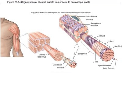
Organization of Skeletal Muscle
Skeletal muscle is organized from the macroscopic to microscopic levels, allowing for efficient contraction and force generation. The structure includes several layers of connective tissue and specialized cellular components.
Muscle: An organ composed of muscle tissue, connective tissue, nerves, and blood vessels.
Fascicle: A bundle of muscle fibers surrounded by perimysium.
Muscle fiber: A single muscle cell, enveloped by endomysium.
Myofibril: Long contractile structures within a muscle fiber, composed of repeating sarcomeres.
Epimysium: Connective tissue surrounding the entire muscle.
Perimysium: Connective tissue surrounding each fascicle.
Endomysium: Connective tissue surrounding each individual muscle fiber.
Sarcoplasmic reticulum: Stores and releases calcium ions necessary for muscle contraction.
T-tubule: Invaginations of the sarcolemma that transmit electrical impulses deep into the muscle fiber.
Thin filaments (Actin): Protein filaments that slide during muscle contraction.
Thick filaments (Myosin): Protein filaments that pull actin during contraction.
Mnemonic: Epi = Entire muscle, Peri = Fascicle, Endo = Individual muscle fiber.
Types of Muscle Tissue
Muscle tissue is classified into three types based on structure, function, and location.
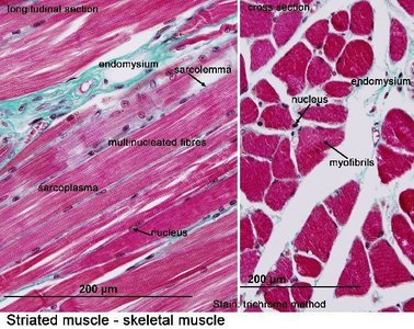
Skeletal Muscle: Voluntary, striated, attached to bones, multinucleated.
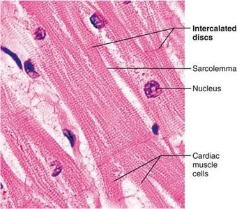
Cardiac Muscle: Involuntary, striated, found only in the heart, branching cells with intercalated discs.
Smooth Muscle: Involuntary, nonstriated, found in walls of organs and blood vessels, spindle-shaped cells.
Supportive Connective Tissues
Connective tissues provide structural support and flexibility to the body.
Hyaline cartilage: Smooth, glassy cartilage found in the nose, trachea, and ends of bones.
Fibrocartilage: Strong cartilage that absorbs shock, found in intervertebral discs and pubic symphysis.
Elastic cartilage: Flexible cartilage found in the external ear and epiglottis.
Bone: Hard connective tissue that supports and protects the body.
Neuromuscular Junctions (NMJ)
The neuromuscular junction is the site where a motor neuron communicates with a muscle fiber to initiate contraction.
Neuromuscular junction: Synapse between a motor neuron and a muscle fiber.
Motor end plate: Specialized region of the muscle fiber membrane that receives the nerve signal.
Acetylcholine (ACh): Neurotransmitter released by the motor neuron to stimulate muscle contraction.
Pectoral Girdle
Clavicle and Scapula
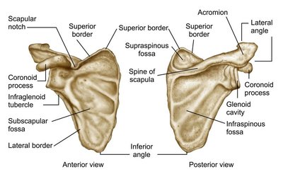
The pectoral girdle connects the upper limbs to the axial skeleton and consists of the clavicle and scapula.
Clavicle: Sternal end (medial, attached to sternum), acromial end (lateral, attached to scapula).
Scapula: Supraspinous fossa, infraspinous fossa, spine, acromion, coracoid process, glenoid cavity/fossa.
Upper Limbs
Humerus
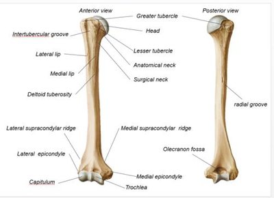
The humerus is the bone of the upper arm, articulating with the scapula and forearm bones.
Head: Rounded proximal end; fits into the glenoid cavity.
Greater tubercle: Large lateral projection near the head.
Lesser tubercle: Smaller anterior projection near the head.
Deltoid tuberosity: Roughened area on the shaft for deltoid attachment.
Trochlea: Spool-shaped distal surface; articulates with the ulna.
Capitulum: Rounded distal surface; articulates with the radius.
Medial/Lateral epicondyle: Distal projections for muscle attachment.
Radius and Ulna
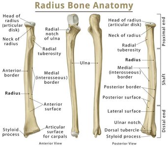
The radius and ulna are the bones of the forearm, connected by the interosseous membrane.
Radius: Head (proximal), tuberosity, styloid process (distal).
Ulna: Trochlear notch, olecranon, coronoid process, styloid process.
Interosseous membrane: Fibrous connective tissue connecting radius and ulna.
Carpal, Metacarpal, and Phalangeal Bones
The wrist and hand are composed of carpal bones, metacarpals, and phalanges.
Carpal bones: Eight bones forming the wrist.
Metacarpals: Five bones of the palm, numbered I–V from thumb to pinky.
Phalanges: Proximal, middle, and distal sections; thumb has only proximal and distal phalanges.
Pelvic Girdle
Coxal Bones and Pelvic Structures
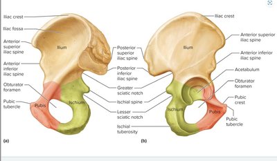
The pelvic girdle supports the lower limbs and consists of the coxal bones, which are fused from three bones.
Ilium: Large, upper flared portion.
Ischium: Lower posterior portion; supports body weight when sitting.
Pubis: Anterior portion.
Pubic symphysis: Cartilaginous joint connecting left and right pubic bones.
Pelvic brim, inlet, outlet: Boundaries and openings of the pelvis.
Acetabulum: Socket for the femoral head.
Obturator foramen: Large opening formed by pubis and ischium.
Lower Limbs
Femur and Patella
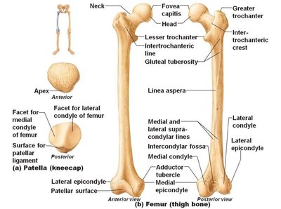
The femur is the thigh bone, the longest bone in the body, and the patella is the kneecap.
Femur: Head, fovea capitis, greater/lesser trochanter, linea aspera, condyles, epicondyles, intercondylar fossa, patellar surface.
Patella: Largest sesamoid bone, protects knee joint, improves quadriceps leverage.
Tibia and Fibula
The tibia and fibula are the bones of the lower leg, forming the ankle joint with the tarsal bones.
Tibia: Medial/lateral condyle, intercondylar eminence, medial malleolus.
Fibula: Head, lateral malleolus.
Tarsal, Metatarsal, and Phalangeal Bones
The foot is composed of tarsal bones, metatarsals, and phalanges.
Tarsal bones: Seven bones, including calcaneus (heel) and talus (ankle).
Metatarsals: Five bones, numbered I–V from big toe to little toe.
Phalanges: Proximal, middle, distal; big toe has only proximal and distal phalanges.
Vertebral Column
Vertebrae Types and Features
The vertebral column consists of cervical, thoracic, lumbar, sacral, and coccygeal vertebrae, each with distinct features.
Cervical: 7 vertebrae (neck).
Thoracic: 12 vertebrae (upper/middle back).
Lumbar: 5 vertebrae (lower back).
Sacrum: Usually 5 fused vertebrae.
Coccyx: Usually 4 fused vertebrae (tailbone).
Mnemonic: 7-12-5 = Cervical, Thoracic, Lumbar.
Special Cervical Vertebrae: Atlas (C1) and Axis (C2)
The atlas and axis are specialized cervical vertebrae that allow for head movement.
Atlas (C1): Supports the skull, allows nodding "yes", lacks vertebral body.
Axis (C2): Has the dens (odontoid process), allows rotation of the head ("no").
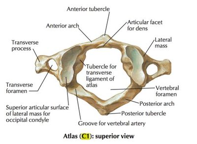
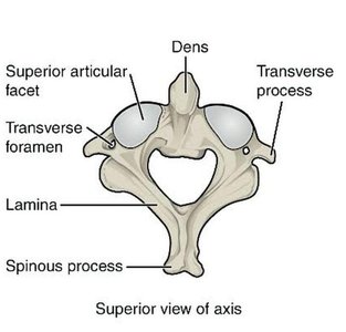
Vertebral Features
Body: Weight-bearing anterior portion.
Vertebral foramen: Opening for the spinal cord.
Vertebral arch: Posterior part of the vertebral foramen.
Spinous process: Posterior projection.
Transverse process: Lateral projections.
Superior/inferior articular processes: Articulate with adjacent vertebrae.
Articular facets: Smooth surfaces at articular processes.
Transverse foramen: Opening in cervical vertebrae for blood vessels.
Thoracic Cage
Sternum and Ribs
The thoracic cage protects vital organs and consists of the sternum and ribs.
Sternum: Manubrium (upper), body (middle), xiphoid process (inferior tip).
Ribs: 12 pairs (24 ribs): True ribs (1–7, direct attachment), false ribs (8–12, indirect/no attachment), floating ribs (11–12, no anterior attachment).
Types of Joints
Fibrous, Cartilaginous, and Synovial Joints
Joints are classified by the material connecting bones and their movement capabilities.
Fibrous Joints: Dense fibrous connective tissue; includes sutures, syndesmoses, gomphoses.
Cartilaginous Joints: Cartilage connects bones; includes synchondrosis (hyaline cartilage), symphysis (fibrocartilage).
Synovial Joints: Freely movable, characterized by a joint cavity and synovial fluid; includes gliding, hinge, pivot, condylar, saddle, ball-and-socket types.
Joint Movements
Types of Movements
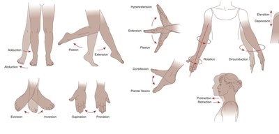
Joints allow for a variety of movements essential for daily activities and locomotion.
Flexion: Decreases the angle between bones.
Extension: Increases the angle between bones.
Abduction: Moves away from the body's midline.
Adduction: Moves toward the body's midline.
Circumduction: Circular, cone-shaped movement.
Rotation: Bone turns around its own axis.
Pronation: Forearm rotates palm downward/posteriorly.
Supination: Forearm rotates palm upward/anteriorly.
Eversion: Sole of foot turns outward.
Inversion: Sole of foot turns inward.
Dorsiflexion: Toes move upward toward the shin.
Plantar flexion: Toes point downward.
Depression: Moves a body part downward.
Elevation: Moves a body part upward.
Memory Tricks: Abduction = Away, Adduction = Add back, Supination = Holding a bowl of soup, Dorsiflexion = Toes up, Plantar flexion = Toes down.
Basic Structural Components of a Synovial Joint
Synovial Joint Anatomy
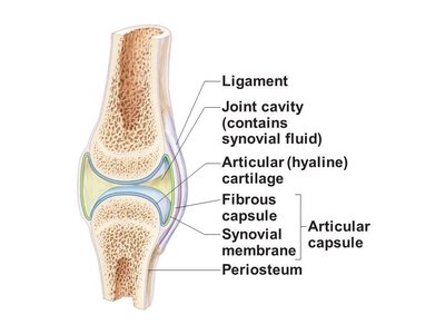
Synovial joints are characterized by a joint cavity, synovial fluid, and specialized structures for movement and shock absorption.
Joint capsule: Encloses and protects the joint.
Menisci: Fibrocartilage pads that improve fit and absorb shock.
Bursa: Small fluid-filled sac that reduces friction.
Ligaments: Connect bone to bone.
Synovial membrane: Inner lining of joint capsule that produces synovial fluid.
Synovial fluid: Lubricates the joint and reduces friction.
Articular cartilage: Covers the ends of bones to reduce friction.
Important Difference: Ligaments connect bone to bone; tendons connect muscle to bone.
Skull Sutures
Major Sutures of the Skull
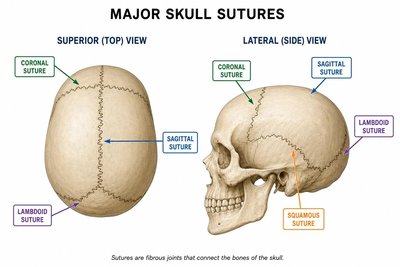
Skull sutures are fibrous joints that connect the bones of the skull, providing stability and growth during development.
Sagittal: Between the two parietal bones.
Coronal: Between the frontal and parietal bones.
Lambdoid: Between the parietal and occipital bones.
Squamous: Between the parietal and temporal bones.
Cranial Bones
Eight Cranial Bones and Their Features
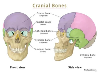
The cranial bones protect the brain and form the structure of the skull.
Frontal Bone: Forms the forehead and roof of the eye sockets; supraorbital foramen/notch, supraorbital margin.
Parietal Bone: Forms the upper and sides of the skull; two parietal bones.
Occipital Bone: Foramen magnum, occipital condyles.
Temporal Bone: Zygomatic process, mandibular fossa, external/internal auditory meatus, styloid process, mastoid process, carotid canal, jugular foramen.
Sphenoid Bone: Optic canal/foramen, superior orbital fissure, sella turcica, hypophyseal fossa.
Ethmoid Bone: Perpendicular plate, cribriform plate, crista galli, ethmoid sinus.
Facial Bones
Fourteen Facial Bones and Their Features
The facial bones form the structure of the face, support teeth, and provide attachment points for muscles.
Maxilla: Upper jaw, palatine processes, maxillary sinuses, alveoli, alveolar processes/margins, infraorbital foramen.
Palatine bone: Forms the posterior hard palate.
Zygomatic bone: Cheekbone.
Lacrimal bone: Medial wall of eye socket, lacrimal groove/fossa.
Nasal bone: Bridge of the nose.
Vomer: Inferior/posterior nasal septum.
Inferior nasal concha: Curved bone inside nasal cavity.
Mandible: Lower jaw, body, ramus; only movable bone of the skull.
Sinus Cavities
Locations and Functions of Sinuses
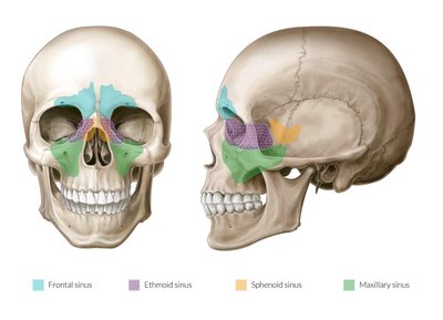
Sinus cavities are air-filled spaces within the skull bones that lighten the skull and aid in voice resonance.
Frontal sinus: Located in the forehead region.
Maxillary sinus: Located in the cheek region.
Sphenoid sinus: Located deep behind the nasal cavity.
Ethmoid sinus: Located between the eyes.
Bone Shapes
Classification of Bones by Shape
Bones are classified by their shape, which relates to their function and location in the body.
Flat bones: Thin, flattened bones for protection (e.g., sternum, parietal bone).
Long bones: Longer than wide, for movement (e.g., femur, humerus).
Short bones: Cube-shaped, for stability (e.g., carpals, tarsals).
Irregular bones: Complex shapes (e.g., vertebrae).
Sesamoid bones: Develop within tendons (e.g., patella).
Sutural bones: Small accessory bones within skull sutures (Wormian bones).
