 Indietro
Anatomy & Physiology Study Guide: Tissues, Connective Tissue, Muscle, Nervous Tissue, and Membranes
Guida di studio - Note intelligenti
 Appunti personalizzati basati sui tuoi materiali, ampliati con definizioni chiave, esempi e contesto.
Tissues: The Living Fabric
Overview of Tissue Types
Tissues are groups of cells with similar structure and function. The four primary tissue types in the human body are epithelial, connective, muscle, and nervous tissues. This guide focuses on connective, muscle, and nervous tissues, as well as covering and lining membranes.
Connective Tissue
Connective Tissue Basics & Characteristics
Connective tissue is the most abundant and widely distributed tissue in the body. It serves multiple functions, including binding and support, protection, insulation, storage of reserve fuel, and transportation of substances (such as blood).
Common embryonic origin: All connective tissues arise from mesenchyme.
Varying degrees of vascularity: Cartilage is avascular, bone is highly vascularized.
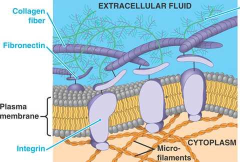
Extracellular matrix (ECM): Cells are suspended in a protein-sugar mesh that supports cells, allowing them to bear weight and withstand tension.
Main Classes of Connective Tissue
Connective tissue proper
Cartilage
Bone
Blood
Structural Elements of Connective Tissue
All connective tissues have three main elements:
Ground substance: Unstructured gel-like material that fills space between cells and allows solutes to diffuse between blood capillaries and cells.
Fibers: Provide support and strength. Three types: collagen, elastic, and reticular.
Cells: Include "blast" (immature, matrix-secreting) and "cyte" (mature, matrix-maintaining) cells, as well as fat cells, white blood cells, mast cells, and macrophages.

Comparison of Classes of Connective Tissues
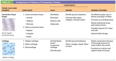
The following tables summarize the main subclasses, cell types, matrix components, and general features of connective tissues:
Tissue Class and Example | Subclasses | Cells | Matrix | General Features |
|---|---|---|---|---|
Connective Tissue Proper | Loose (Areolar, Adipose, Reticular); Dense (Regular, Irregular, Elastic) | Fibroblasts, Defense cells, Adipocytes | Gel-like ground substance; All three fiber types: collagen, reticular, elastic | Six different types; functions as binding tissue; resists mechanical stress; provides reservoir for water and salts; nutrient (fat) storage |
Cartilage | Hyaline, Elastic, Fibrocartilage | Chondroblasts (in growing cartilage), Chondrocytes | Gel-like ground substance; fibers: collagen, elastic fibers in some | Resists compression; functions to cushion and support body structures |

Tissue Class and Example | Subclasses | Cells | Matrix | General Features |
|---|---|---|---|---|
Bone Tissue | Compact bone, Spongy bone | Osteoblasts, Osteocytes | Gel-like ground substance calcified with inorganic salts; fibers: collagen | Hard tissue that resists both compression and tension; functions in support |
Blood | See Chapter 17 for details | Erythrocytes (RBC), Leukocytes (WBC), Platelets | Plasma; no fibers | Fluid tissue; functions to carry O2, CO2, nutrients, wastes, and other substances |
Connective Tissue Proper
Loose Connective Tissue
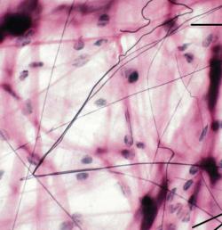
Areolar: Most widely distributed; supports and binds other tissues; contains all fiber types; acts as water reservoir.
Adipose: Stores nutrients; shock absorption, insulation, energy storage; cells are adipocytes.
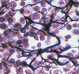
Reticular: Mesh-like stroma supports blood cells in lymph nodes, spleen, and bone marrow; contains reticular fibers and reticular cells.
Dense Connective Tissue
Dense regular: Parallel collagen fibers; high tensile strength; found in tendons and ligaments.
Dense irregular: Thicker, irregularly arranged collagen fibers; resists tension from many directions; found in dermis and fibrous capsules.
Elastic: Contains many elastic fibers; found in walls of large arteries and certain ligaments.
Cartilage
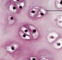
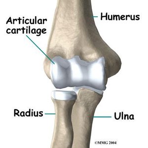
Hyaline cartilage: Most abundant; provides support and flexibility; found at tips of long bones, nose, trachea, larynx, and ribs.
Elastic cartilage: More elastic fibers; found in ears and epiglottis.
Fibrocartilage: Strong; found in intervertebral discs and knee.
Bone (Osseous Tissue)
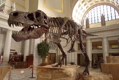
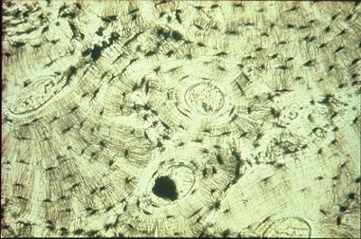
Bones support and protect body structures, store fat, and synthesize blood cells. They have more collagen than cartilage and contain inorganic calcium salts. Osteoblasts produce matrix, osteocytes maintain it, and both reside in lacunae. Bone is highly vascularized and heals well.
Blood
Blood is a fluid connective tissue consisting of cells (red and white blood cells, platelets) surrounded by plasma. It functions in transport of gases, nutrients, wastes, and other substances.
Muscle Tissue
Types of Muscle Tissue
Muscle tissue is highly vascularized and responsible for movement. Muscle cells contain myofilaments made of actin and myosin, which enable contraction. There are three types:
Skeletal muscle: Voluntary, striated, multinucleate; attached to bones.
Cardiac muscle: Involuntary, striated, one nucleus per cell, intercalated discs; found only in heart.
Smooth muscle: Involuntary, non-striated, spindle-shaped cells; found in walls of hollow organs.
Nervous Tissue
Structure and Function
Nervous tissue is the main component of the nervous system (brain, spinal cord, nerves). It regulates and controls body functions. It consists of:
Neurons: Specialized cells that generate and conduct nerve impulses.
Supporting cells: Support, insulate, and protect neurons.
Covering and Lining Membranes
Types and Characteristics
Membranes are composed of at least two primary tissue types: an epithelium bound to underlying connective tissue proper. Three types:
Cutaneous membrane: Skin; keratinized stratified squamous epithelium attached to connective tissue (dermis); dry membrane.
Mucous membrane: Lines body cavities open to exterior (digestive, respiratory, urogenital tracts); moist, may secrete mucus.
Serous membrane: Lines closed ventral body cavities; constructed from simple squamous epithelium resting on areolar connective tissue; parietal serosae line cavity walls, visceral serosae cover organs; cavity between layers filled with serous fluid.
Tissue Repair
Regeneration vs. Fibrosis
When tissue is injured, repair occurs via two major mechanisms:
Regeneration: Same kind of tissue replaces destroyed tissue; original function restored.
Fibrosis: Connective tissue replaces destroyed tissue; original function lost (scar tissue).
Steps in Tissue Repair
Inflammation: Release of inflammatory chemicals, dilation of blood vessels, increased permeability, clotting.
Organization: Blood clot replaced with granulation tissue; epithelium regenerates; fibroblasts produce collagen fibers; debris is phagocytized.
Regeneration and fibrosis: Scab detaches; epithelium thickens; results in fully regenerated epithelium with underlying scar tissue.
Regenerative Capacity of Different Tissues
High capacity: Epithelial tissues, bone, areolar connective tissue, dense irregular connective tissue, blood-forming tissue.
Moderate capacity: Smooth muscle, dense regular connective tissue.
Low/no capacity: Cardiac muscle, nervous tissue of brain and spinal cord.
Aging and Tissues
As the body ages, epithelia thin, tissue repair becomes less efficient, bone and muscle tissues atrophy, and DNA mutations increase cancer risk.
