 Indietro
Anatomy & Physiology Unit 1 Exam Study Guide – Step-by-Step Guidance
Guida di studio - Note intelligenti
 Appunti personalizzati basati sui tuoi materiali, ampliati con definizioni chiave, esempi e contesto.
Q1. What are the hormones of the adrenal gland and their functions?
Background
Topic: Endocrine System – Adrenal Gland
This question tests your knowledge of the hormones produced by the adrenal cortex and medulla, and their physiological roles in the body.
Key Terms and Concepts:
Adrenal cortex: Produces corticosteroids (e.g., cortisol, aldosterone, and androgens).
Adrenal medulla: Produces catecholamines (e.g., epinephrine and norepinephrine).
Step-by-Step Guidance
Identify the two main regions of the adrenal gland and the types of hormones each produces.
List the major hormones from the adrenal cortex and describe their primary functions (e.g., regulation of metabolism, stress response, electrolyte balance).
List the hormones from the adrenal medulla and explain their role in the fight-or-flight response.
Consider how these hormones interact with other body systems to maintain homeostasis.
Try solving on your own before revealing the answer!
Final Answer:
The adrenal cortex produces cortisol (regulates metabolism and stress response), aldosterone (controls sodium and potassium balance), and androgens (sex hormones). The adrenal medulla produces epinephrine and norepinephrine, which increase heart rate, blood pressure, and blood glucose during stress (fight-or-flight response).
Q2. What are the anatomical and physiological connections between the hypothalamus and pituitary glands?
Background
Topic: Endocrine System – Hypothalamic-Pituitary Axis
This question examines your understanding of how the hypothalamus communicates with the anterior and posterior pituitary glands to regulate hormone secretion.
Key Terms and Concepts:
Hypothalamus: Brain region controlling the pituitary gland.
Anterior pituitary (adenohypophysis): Receives releasing/inhibiting hormones via the hypophyseal portal system.
Posterior pituitary (neurohypophysis): Releases hormones produced in the hypothalamus via nerve fibers.
Step-by-Step Guidance
Describe the physical connection between the hypothalamus and pituitary gland (infundibulum).
Explain how the hypothalamus controls the anterior pituitary using the portal blood system.
Discuss how the hypothalamus sends hormones directly to the posterior pituitary via axons.
Identify examples of hormones involved in each pathway.
Try solving on your own before revealing the answer!
Final Answer:
The hypothalamus connects to the anterior pituitary via the hypophyseal portal system (blood vessels) and to the posterior pituitary via nerve fibers. It regulates the anterior pituitary by secreting releasing/inhibiting hormones and sends oxytocin and ADH to the posterior pituitary for release.
Q3. What are the functional classifications of hormones?
Background
Topic: Endocrine System – Hormone Classification
This question tests your ability to categorize hormones based on their chemical structure and mechanism of action.
Key Terms and Concepts:
Peptide hormones: Made of amino acids, water-soluble.
Steroid hormones: Derived from cholesterol, lipid-soluble.
Amino acid derivatives: Modified amino acids (e.g., thyroid hormones, catecholamines).
Step-by-Step Guidance
List the main chemical classes of hormones (peptide, steroid, amino acid derivatives).
Describe the solubility and receptor location for each class (membrane-bound vs. intracellular).
Give examples of hormones in each class.
Explain how the mechanism of action differs between classes.
Try solving on your own before revealing the answer!
Final Answer:
Hormones are classified as peptide/protein hormones (e.g., insulin), steroid hormones (e.g., cortisol), and amino acid derivatives (e.g., epinephrine, thyroid hormones). Peptide hormones act on cell surface receptors, steroid hormones act on intracellular receptors, and amino acid derivatives can act on either.
Q4. What hormones are involved in calcium homeostasis?
Background
Topic: Endocrine System – Calcium Regulation
This question focuses on the hormones that regulate blood calcium levels and their effects on bones, kidneys, and intestines.
Key Terms and Concepts:
Parathyroid hormone (PTH): Increases blood calcium.
Calcitonin: Lowers blood calcium.
Calcitriol (active vitamin D): Increases calcium absorption from the gut.
Step-by-Step Guidance
Identify the glands that secrete each hormone involved in calcium homeostasis.
Describe the effect of each hormone on blood calcium levels.
Explain the target organs and actions (bone, kidney, intestine) for each hormone.
Consider the feedback mechanisms that regulate these hormones.
Try solving on your own before revealing the answer!
Final Answer:
Parathyroid hormone (PTH) increases blood calcium by stimulating bone resorption, increasing kidney reabsorption, and activating vitamin D. Calcitonin lowers blood calcium by inhibiting bone resorption. Calcitriol increases calcium absorption from the intestines.
Q5. What is the function of the thymus gland?
Background
Topic: Endocrine and Immune Systems – Thymus Gland
This question tests your understanding of the thymus gland’s role in immune system development, especially in children.
Key Terms and Concepts:
Thymus: Lymphoid organ in the mediastinum.
T lymphocytes (T cells): White blood cells essential for adaptive immunity.
Step-by-Step Guidance
Locate the thymus gland anatomically and describe its structure.
Explain the role of the thymus in T cell maturation and immune function.
Discuss how the thymus changes with age.
Try solving on your own before revealing the answer!
Final Answer:
The thymus gland is responsible for the maturation of T lymphocytes, which are crucial for adaptive immunity. It is most active during childhood and shrinks after puberty.
Q6. How is blood sugar homeostasis maintained?
Background
Topic: Endocrine System – Glucose Regulation
This question examines your knowledge of the hormones and feedback mechanisms that regulate blood glucose levels.
Key Terms and Concepts:
Insulin: Lowers blood glucose by promoting uptake into cells.
Glucagon: Raises blood glucose by stimulating glycogen breakdown.
Negative feedback: Maintains homeostasis.
Step-by-Step Guidance
Identify the pancreatic cells that secrete insulin and glucagon.
Describe the effects of insulin and glucagon on blood glucose levels.
Explain the feedback mechanisms that regulate hormone secretion in response to blood glucose changes.
Try solving on your own before revealing the answer!
Final Answer:
Blood sugar homeostasis is maintained by insulin (from beta cells, lowers glucose) and glucagon (from alpha cells, raises glucose). Negative feedback ensures that hormone secretion adjusts to keep glucose levels stable.
Q7. Describe the anatomy of the heart, including the conduction system.
Background
Topic: Cardiovascular System – Heart Anatomy and Physiology
This question tests your understanding of the heart’s chambers, valves, and the electrical conduction system that coordinates heartbeats.
Key Terms and Concepts:
Chambers: Right/left atria, right/left ventricles.
Valves: Tricuspid, pulmonary, mitral, aortic.
Conduction system: SA node, AV node, bundle of His, bundle branches, Purkinje fibers.
Step-by-Step Guidance
Identify the four chambers and four valves of the heart.
Describe the flow of blood through the heart chambers and valves.
Outline the pathway of electrical conduction from the SA node to the Purkinje fibers.
Explain how the conduction system coordinates contraction of the heart chambers.
Try solving on your own before revealing the answer!
Final Answer:
The heart has four chambers (right/left atria and ventricles) and four valves (tricuspid, pulmonary, mitral, aortic). The conduction system starts at the SA node, travels to the AV node, bundle of His, bundle branches, and Purkinje fibers, ensuring coordinated contraction.
Q8. How does blood flow through the chambers of the heart?
Background
Topic: Cardiovascular System – Cardiac Circulation
This question focuses on the sequence of blood flow through the heart’s chambers and valves.
Key Terms and Concepts:
Right atrium, right ventricle, left atrium, left ventricle.
Valves: Tricuspid, pulmonary, mitral, aortic.
Step-by-Step Guidance
Trace the path of deoxygenated blood entering the right atrium from the body.
Follow the blood through the right ventricle and into the pulmonary circulation.
Describe the return of oxygenated blood to the left atrium and its passage to the left ventricle.
Explain how blood is pumped from the left ventricle to the systemic circulation.
Try solving on your own before revealing the answer!
Final Answer:
Blood flows from the body into the right atrium, through the tricuspid valve to the right ventricle, then to the lungs via the pulmonary valve. Oxygenated blood returns to the left atrium, passes through the mitral valve to the left ventricle, and is pumped to the body via the aortic valve.
Q9. Describe the anatomy of the walls of the heart.
Background
Topic: Cardiovascular System – Heart Wall Structure
This question tests your knowledge of the three layers of the heart wall and their functions.
Key Terms and Concepts:
Epicardium: Outer layer.
Myocardium: Middle, muscular layer.
Endocardium: Inner lining.
Step-by-Step Guidance
List the three layers of the heart wall from outermost to innermost.
Describe the composition and function of each layer.
Explain the importance of the myocardium in heart contraction.
Try solving on your own before revealing the answer!
Final Answer:
The heart wall consists of the epicardium (outer protective layer), myocardium (thick muscular layer responsible for contraction), and endocardium (inner lining that contacts blood).
Q10. What is the anatomy of the coronary cardiac circuit?
Background
Topic: Cardiovascular System – Coronary Circulation
This question examines your understanding of the blood vessels that supply the heart muscle itself.
Key Terms and Concepts:
Coronary arteries: Right and left, branch from the aorta.
Cardiac veins: Drain deoxygenated blood from the myocardium.
Coronary sinus: Collects blood from cardiac veins and returns it to the right atrium.
Step-by-Step Guidance
Identify the main coronary arteries and their branches.
Describe the pathway of blood through the coronary arteries to the myocardium.
Explain how deoxygenated blood is collected by cardiac veins and returned to the right atrium via the coronary sinus.
Try solving on your own before revealing the answer!
Final Answer:
The coronary circuit includes the right and left coronary arteries (from the aorta), their branches, cardiac veins, and the coronary sinus, which returns deoxygenated blood to the right atrium.
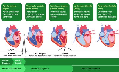
Q11. What are the four phases of the cardiac cycle?
Background
Topic: Cardiovascular System – Cardiac Cycle
This question tests your knowledge of the sequence of mechanical and electrical events in one heartbeat.
Key Terms and Concepts:
Atrial systole, ventricular systole, ventricular diastole (early and late).
Contraction and relaxation phases.
Step-by-Step Guidance
List the four main phases of the cardiac cycle.
Describe what happens in the atria and ventricles during each phase.
Relate each phase to the opening and closing of heart valves.
Connect the phases to the corresponding ECG waves.
Try solving on your own before revealing the answer!
Final Answer:
The four phases are: atrial systole (atria contract), ventricular systole (ventricles contract), early ventricular diastole (ventricles relax, semilunar valves close), and late ventricular diastole (chambers fill passively). Each phase corresponds to specific ECG waves and valve movements.
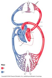
Q12. Describe the pulmonary and systemic blood circulation.
Background
Topic: Cardiovascular System – Circulatory Pathways
This question tests your understanding of how blood circulates through the lungs (pulmonary) and the rest of the body (systemic).
Key Terms and Concepts:
Pulmonary circulation: Right ventricle to lungs to left atrium.
Systemic circulation: Left ventricle to body to right atrium.
Step-by-Step Guidance
Trace the path of deoxygenated blood from the right ventricle to the lungs and back to the left atrium (pulmonary circuit).
Trace the path of oxygenated blood from the left ventricle to the body and back to the right atrium (systemic circuit).
Identify the major vessels involved in each circuit.
Explain the purpose of each circuit in oxygenating blood and delivering nutrients.
Try solving on your own before revealing the answer!
Final Answer:
Pulmonary circulation carries deoxygenated blood from the right ventricle to the lungs and returns oxygenated blood to the left atrium. Systemic circulation carries oxygenated blood from the left ventricle to the body and returns deoxygenated blood to the right atrium.
