 Indietro
Anatomy of the Muscular System: Muscles of the Upper and Lower Limbs
Guida di studio - Note intelligenti
 Appunti personalizzati basati sui tuoi materiali, ampliati con definizioni chiave, esempi e contesto.
Anatomy of the Muscular System
Muscles Crossing the Shoulder and Elbow Joints
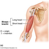
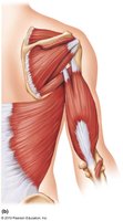
The muscles that cross the shoulder and elbow joints are essential for movements of the upper limb, including flexion, extension, and rotation. These muscles originate from the scapula, humerus, or other bones and insert onto the forearm bones, enabling complex actions.
Biceps brachii: Originates from the coracoid process and glenoid cavity, inserts on the radius. It is located anteriorly and functions to flex the elbow joint and supinate the forearm. It is a weak flexor of the arm at the shoulder.
Brachialis: Originates from the humerus and inserts on the ulna. It lies immediately deep to the biceps brachii and is the major forearm flexor, lifting the ulna as the biceps lifts the radius.
Brachioradialis: Originates from the humerus and inserts on the radius. It is superficial and acts as a synergist in forearm flexion, especially when the forearm is semi-pronated.
Triceps brachii: Originates from the scapula and humerus, inserts on the ulna. It is the only muscle of the posterior compartment of the arm and is a powerful forearm extensor. It may also help stabilize the shoulder joint.
Muscles of the Forearm: Movements of the Wrist, Hand, and Fingers
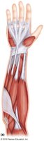
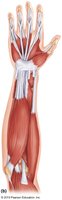
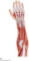
Forearm muscles are divided into two functional groups: those that move the wrist and those that move the fingers and thumb. These muscles allow for a wide range of precise movements necessary for daily activities.
Pronator teres: Pronates the forearm and is a weak flexor of the elbow.
Flexor carpi radialis: Powerful flexor of the wrist and abducts the hand.
Flexor carpi ulnaris: Powerful flexor of the wrist, adducts the hand in concert with extensor carpi ulnaris, and stabilizes the wrist during finger extension.
Flexor digitorum superficialis: Flexes the wrist and middle phalanges of fingers 2-5.
Extensor carpi radialis longus/brevis: Extends and abducts the wrist.
Extensor digitorum: Prime mover of finger extension, extends the wrist, and can abduct fingers.
Supinator: Assists biceps brachii in supinating the forearm.
Muscles of the Hip, Thigh, and Leg
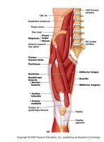
The muscles of the hip, thigh, and leg are responsible for movements such as walking, running, and jumping. They include flexors, extensors, adductors, and rotators that act on the hip and knee joints.
Iliopsoas: Composite of two muscles, prime mover of hip flexion.
Sartorius: Strap-like, longest muscle; flexes and laterally rotates thigh, weak knee flexor.
Adductors (magnus, longus, brevis): Adduct, flex, and medially rotate the thigh.
Rectus femoris: Extends the knee and flexes the thigh at the hip.
Vastus (lateralis, medialis, intermedius): All extend the knee only.
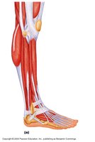
Tibialis anterior: Prime mover of dorsiflexion, inverts the foot.
Extensor digitorum longus: Dorsiflexes the foot, prime mover of toe extension.
Pectineus: Adducts, flexes, and medially rotates the thigh.
Gracilis: Adducts the thigh, medially rotates the leg.
Muscles of the Gluteal Region and Posterior Thigh
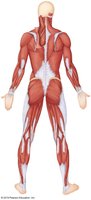
The gluteal muscles and hamstrings are crucial for extension, abduction, and rotation of the thigh, as well as flexion of the knee. These muscles are important for activities such as climbing, running, and maintaining posture.
Gluteus maximus: Major thigh extensor, largest and most superficial buttock muscle.
Gluteus medius: Abducts and medially rotates the thigh, especially during walking.
Hamstrings (biceps femoris, semitendinosus, semimembranosus): All extend the thigh and flex the knee.
Muscles of the Lower Leg: Movements of the Ankle and Foot
Muscles of the lower leg control movements of the ankle and foot, including dorsiflexion, plantar flexion, inversion, and eversion. These muscles are essential for locomotion and balance.
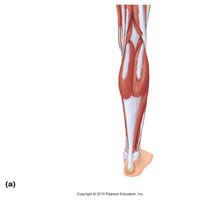
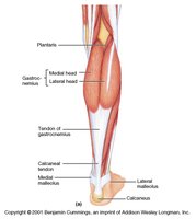
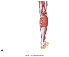
Gastrocnemius: Plantar flexes the foot when the knee is extended, flexes the knee if the foot is dorsiflexed.
Soleus: Deeper muscle, plantar flexes the ankle during walking, running, and dancing.
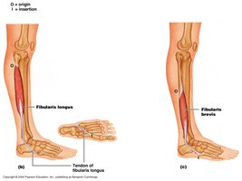
Fibularis (peroneus) longus: Plantar flexes and everts the foot, helps keep the foot flat on the ground.
Calcaneal (Achilles) tendon: Largest tendon in the body, connects gastrocnemius and soleus to the calcaneus.
Summary Table: Major Muscles and Their Actions
The following table summarizes the major muscles discussed and their primary actions:
Muscle | Origin | Insertion | Action |
|---|---|---|---|
Biceps brachii | Coracoid process & glenoid cavity | Radius | Flexes elbow, supinates forearm |
Brachialis | Humerus | Ulna | Forearm flexion |
Triceps brachii | Scapula/humerus | Ulna | Forearm extension |
Flexor carpi radialis | Humerus | Metacarpals | Flexes wrist, abducts hand |
Flexor carpi ulnaris | Humerus/ulna | Metacarpals | Flexes wrist, adducts hand |
Rectus femoris | Iliac spine | Patella & tibia | Extends knee, flexes thigh |
Tibialis anterior | Tibia | Tarsals | Dorsiflexion, inverts foot |
Gastrocnemius | Femur | Calcaneal tendon | Plantar flexion, knee flexion |
Fibularis longus | Fibula | 1st metatarsal | Plantar flexion, eversion |
Key Terms and Definitions
Flexion: Decreasing the angle between two bones, such as bending the elbow.
Extension: Increasing the angle between two bones, such as straightening the knee.
Abduction: Moving a limb away from the midline of the body.
Adduction: Moving a limb toward the midline of the body.
Supination: Rotating the forearm so the palm faces upward.
Pronation: Rotating the forearm so the palm faces downward.
Dorsiflexion: Raising the foot upwards towards the shin.
Plantar flexion: Pointing the foot downward away from the leg.
Example: Application in Daily Activities
Climbing stairs: Requires extension of the thigh by the gluteus maximus and flexion of the knee by the hamstrings.
Writing: Involves fine motor control by the forearm and hand muscles, such as flexor digitorum superficialis and extensor digitorum.
Walking: Utilizes coordinated actions of the iliopsoas, quadriceps, hamstrings, tibialis anterior, and gastrocnemius.
Additional info: Academic context and expanded explanations were added to ensure completeness and clarity for exam preparation.
