 Indietro
Arteries: Structure, Function, and Major Branches
Guida di studio - Note intelligenti
 Appunti personalizzati basati sui tuoi materiali, ampliati con definizioni chiave, esempi e contesto.
Arteries and Blood Vessels
Structure and Function of Arteries
Arteries are blood vessels that carry blood away from the heart. Their elasticity allows them to absorb the pressure waves generated by each heartbeat, while contractility enables them to change diameter, a process regulated by the sympathetic division of the autonomic nervous system (ANS). The contraction of arterial smooth muscle is called vasoconstriction, and relaxation is termed vasodilation, which enlarges the lumen (the interior space of the vessel).
Elasticity: Absorbs pressure waves from the heart.
Contractility: Allows arteries to change diameter.
Vasoconstriction: Contraction of smooth muscle, reducing lumen size.
Vasodilation: Relaxation of smooth muscle, increasing lumen size.
Types of Blood Vessels
Blood vessels are classified based on their function and structure. Arteries carry blood away from the heart, arterioles are the smallest branches leading to capillary beds, and capillaries are the site of exchange between blood and interstitial fluid. Venules collect blood from capillaries, and veins return blood to the heart.
Arteries: Carry blood away from the heart.
Arterioles: Smallest branches of arteries.
Capillaries: Smallest blood vessels; site of exchange.
Venules: Smallest branches of veins.
Veins: Return blood to the heart.
Layers of Vessel Walls
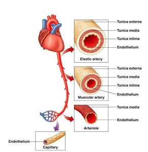
Blood vessel walls consist of three layers: tunica intima, tunica media, and tunica externa. Each layer has distinct structural and functional properties.
Tunica intima: Inner layer; includes endothelial lining, connective tissue, and internal elastic membrane (in arteries).
Tunica media: Middle layer; contains concentric sheets of smooth muscle and external elastic membrane.
Tunica externa: Outer layer; anchors vessel to adjacent tissues, contains collagen and elastic fibers, and vasa vasorum in large vessels.
Differences Between Arteries and Veins
Arteries and veins differ in structure and function. Arteries have thicker walls and higher blood pressure, a small round lumen when constricted, and are more elastic. Veins have a large, irregular lumen, less elasticity, and contain valves to prevent backflow.
Arteries: Thicker walls, higher pressure, more elastic, folded endothelium when constricted.
Veins: Thinner walls, lower pressure, less elastic, large irregular lumen, contain valves.
Types of Arteries
Elastic Arteries (Conducting Arteries)
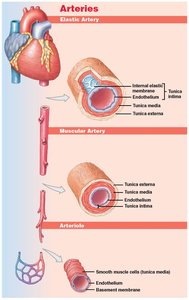
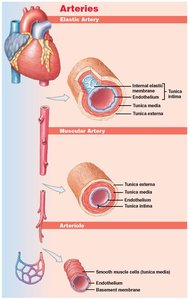
Elastic arteries are large vessels such as the pulmonary trunk and aorta. Their tunica media contains many elastic fibers and few muscle cells, allowing them to even out pulse force and absorb pressure fluctuations.
Examples: Aorta, pulmonary trunk.
Function: Conduct blood from the heart and dampen pressure changes.
Muscular Arteries (Distribution Arteries)
Muscular arteries are medium-sized and distribute blood to various organs. Their tunica media contains many muscle cells, allowing for greater control of blood flow.
Examples: Brachial, radial, femoral arteries.
Function: Distribute blood to specific regions and organs.
Arterioles (Resistance Vessels)
Arterioles are small vessels with little or no tunica externa and a thin or incomplete tunica media. They regulate blood flow into capillary beds and are key in controlling systemic vascular resistance.
Function: Regulate blood flow and pressure.
Major Systemic Arteries and the Aorta
Parts of the Aorta
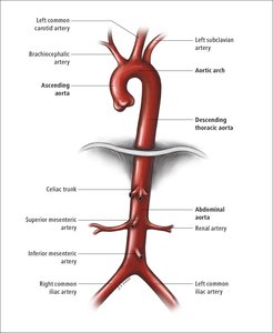
The aorta is the largest artery in the body and is divided into several parts: ascending aorta, aortic arch, descending thoracic aorta, and abdominal aorta. Each part gives rise to important branches supplying various regions.
Ascending aorta: Rises from the left ventricle.
Aortic arch: Curves and gives rise to major branches.
Descending aorta: Continues downward; thoracic and abdominal segments.
Branches of the Aortic Arch
The aortic arch delivers blood to the head, neck, shoulders, and upper limbs. Its main branches are:
Brachiocephalic trunk: Gives rise to right subclavian and right common carotid arteries.
Left common carotid artery: Supplies head and neck.
Left subclavian artery: Supplies arms, chest wall, shoulders, back, and CNS.
Branches of the Abdominal Aorta
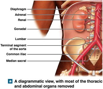
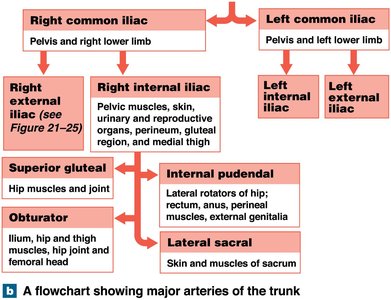
The abdominal aorta divides into the left and right common iliac arteries at its terminal segment. It gives rise to both unpaired branches (to visceral organs) and paired branches (to body wall, kidneys, adrenal glands, gonads, and structures outside the abdominopelvic cavity).
Unpaired branches: Supply visceral organs.
Paired branches: Supply body wall and various organs.
Clinical Conditions: Blood Pressure and Vascular Disorders
Blood Pressure
Blood pressure is the force exerted by circulating blood on the walls of blood vessels. Normal blood pressure is considered to be 120/80 mmHg.
Hypertension: Abnormally high blood pressure (>140/90 mmHg).
Hypotension: Abnormally low blood pressure.
Aneurysm
An aneurysm is a bulge in an arterial wall caused by a weak spot in elastic fibers. If the pressure becomes too great, the vessel may rupture, leading to potentially life-threatening consequences.
Definition: Bulge in arterial wall due to weakness.
Risk: Rupture and hemorrhage.
Summary of Key Points
Arteries carry blood away from the heart.
Three types of arteries: elastic, muscular, and arterioles.
The aorta is divided into ascending, arch, descending (thoracic and abdominal).
Three main branches from the aortic arch.
Abdominal aorta has single and paired branches.
Hypertension and aneurysm are high-risk vascular conditions.
Additional info:
Blood vessel structure is essential for understanding vascular function and pathology.
Regulation of vessel diameter is crucial for maintaining blood pressure and tissue perfusion.
