 Indietro
Articulations and Synovial Joints: Structure, Types, and Movements
Guida di studio - Note intelligenti
 Appunti personalizzati basati sui tuoi materiali, ampliati con definizioni chiave, esempi e contesto.
Articulations (Joints)
Introduction to Joints
Joints, also known as articulations, are points of contact between two or more bones, cartilage and bones, or teeth and bones. The scientific study of joints is called arthrology. Joints serve to hold bones together while permitting movement, playing a crucial role in the skeletal system's function.
Definition: A joint is a location where two or more bones meet.
Functions: Provide stability and allow movement.
Types of contacts: Bone-to-bone, cartilage-to-bone, teeth-to-bone.
Classification of Joints
Joints are classified based on their structure and the degree of movement they allow:
Fibrous Joints: Immovable joints, such as those between cranial bones.
Cartilaginous Joints: Slightly movable joints, such as those between vertebrae.
Synovial Joints: Freely movable joints, such as the knee.
Synovial Joints
Structure of Synovial Joints
Synovial joints are characterized by a synovial cavity that separates the articulating bones, making them freely movable. The structure of a synovial joint includes several key components:
Articular Cartilage: Covers the ends of bones, reduces friction, and absorbs shock.
Articular Capsule: Surrounds the joint; thickenings in the fibrous capsule are called ligaments.
Synovial Membrane: Inner lining of the capsule that secretes synovial fluid.
Synovial Fluid: Contains hyaluronic acid, lubricates the joint, and nourishes articular cartilage.
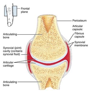
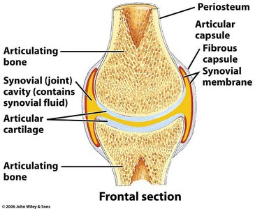
Knee Joint Anatomy
The knee joint is the largest and most complex synovial joint, consisting of three joints within a single synovial cavity. It allows flexion, extension, and slight rotation. The knee is stabilized by accessory ligaments and articular discs (menisci), which increase its stability.
Common injuries: Rupture of the tibial collateral ligament, dislocation of the knee.
Bursae: Fluid-filled sacs that reduce friction; inflammation is called bursitis.
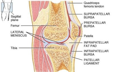
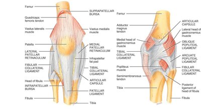
Types of Synovial Joints
Ball-and-Socket Joint
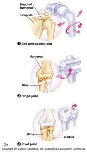
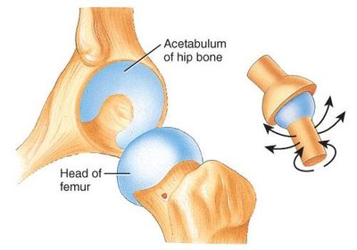
Ball-and-socket joints allow movement in multiple planes, including rotation. The ball fits into a cuplike depression, making these joints multiaxial.
Examples: Shoulder and hip joints.
Movements: Flexion/extension, abduction/adduction, rotation.
Hinge Joint
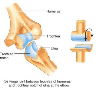
Hinge joints restrict movement to a single plane (uniaxial), similar to a door hinge. The convex surface of one bone fits into the concave surface of another.
Examples: Knee, elbow, ankle, interphalangeal joints.
Movements: Flexion, extension, hyperextension.
Pivot Joint
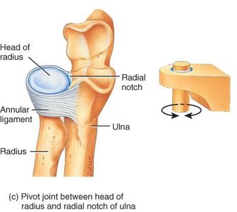
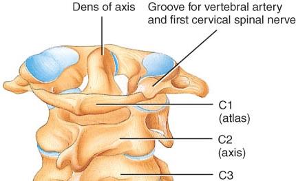
Pivot joints allow rotation around a single axis (monoaxial). A rounded bone surface articulates with a ring formed by another bone and ligament.
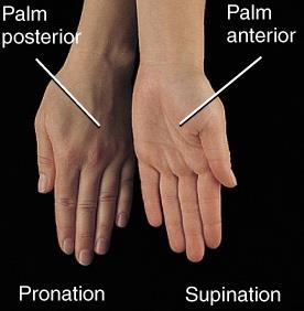
Examples: Proximal radioulnar joint (supination/pronation), atlanto-axial joint (turning head side to side).
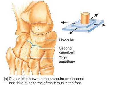
Planar Joint
Planar joints have flat or slightly curved surfaces, allowing only side-to-side gliding movements. Rotation is prevented by ligaments.
Examples: Intercarpal, intertarsal, sternoclavicular, vertebrocostal joints.
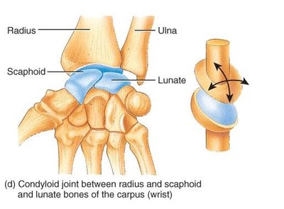
Condyloid (Ellipsoidal) Joint
Condyloid joints feature an oval-shaped projection fitting into an oval depression, allowing biaxial movement (flexion/extension, abduction/adduction).
Examples: Wrist, metacarpophalangeal joints (digits 2-5).
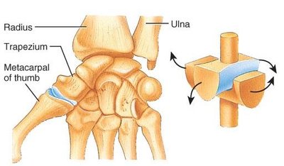
Saddle Joint
Saddle joints have one bone shaped like a saddle and another fitting as a rider. They allow biaxial movement, including circumduction and opposition.
Example: Trapezium of carpus and metacarpal of the thumb.
Factors Affecting Range of Motion at Synovial Joints
Determinants of Joint Movement
The range of motion at synovial joints is influenced by several factors:
Structure and shape of articulating bones
Strength and tautness of ligaments
Arrangement and tension of muscles
Contact of soft parts
Hormones
Disuse
Aging affects joints by decreasing synovial fluid production, thinning articular cartilage, and reducing ligament flexibility.
Homeostatic Imbalances of Joints
Osteoarthritis
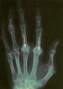
Osteoarthritis is a degenerative joint disease characterized by deterioration of articular cartilage, primarily affecting weight-bearing joints. It is noninflammatory and produces bone spurs that restrict movement.
Symptoms: Pain upon awakening, which disappears with movement.
Rheumatoid Arthritis
Rheumatoid arthritis is an autoimmune disorder where cartilage is attacked, leading to inflammation, swelling, pain, and eventual fusion of the joint.
Other Joint Disorders
Lyme Disease: Bacterial disease transmitted by deer ticks, causing joint stiffness and other symptoms.
Ankylosing Spondylitis: Affects joints between vertebrae and sacrum/hip bone; cause is unknown.
Gouty Arthritis: Sodium urate crystals deposit in joints, causing inflammation and pain; untreated cases may lead to bone fusion.
Sprain vs. Strain
Sprain: Twisting of a joint that stretches or tears ligaments without dislocation; may damage blood vessels, muscles, or tendons.
Strain: Less serious injury; overstretched or partially torn muscle.
Types of Movement at Synovial Joints
Gliding Movements
Gliding movements occur when flat bone surfaces move back and forth or side to side, with no significant change in the angle between bones. These movements are typical of planar joints.
Angular Movements
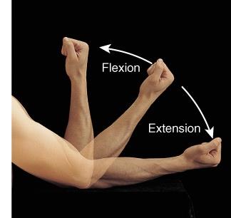
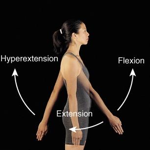
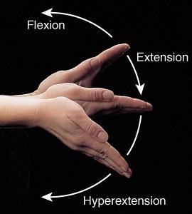
Angular movements involve changes in the angle between articulating bones:
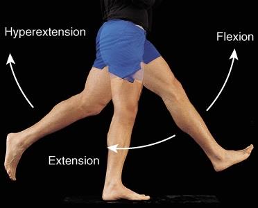
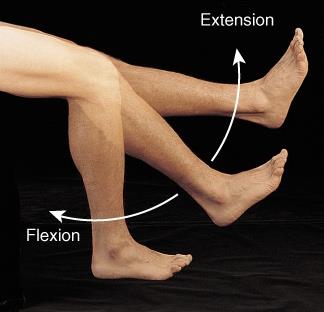
Flexion: Decreases the angle between bones.
Extension: Increases the angle between bones.
Hyperextension: Extension beyond the anatomical position.
Lateral Flexion: Movement of the trunk sideways at the waist.
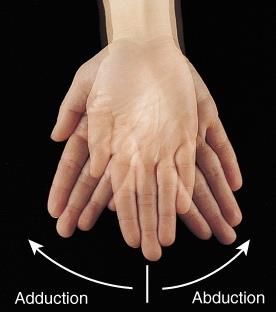
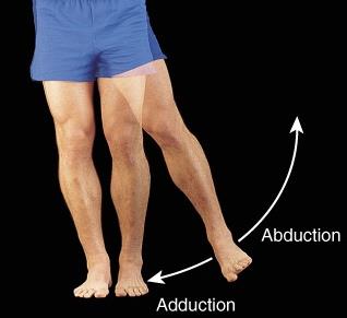
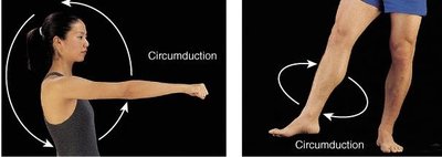
Abduction, Adduction, and Circumduction
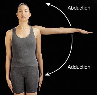
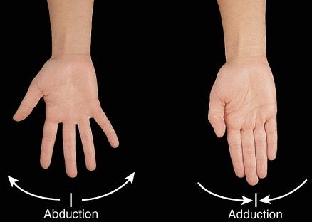
Abduction: Movement of a bone away from the midline.
Adduction: Movement of a bone toward the midline.
Circumduction: Movement of the distal end of a body part in a circle, combining flexion, abduction, extension, and adduction.
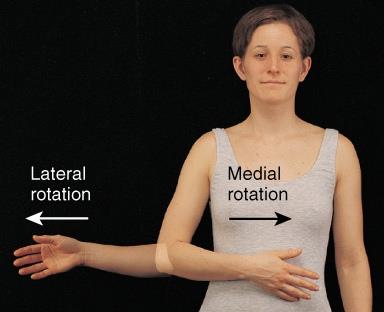
Rotation
Rotation occurs when a bone revolves around its own longitudinal axis. Medial rotation turns the anterior surface toward the midline, while lateral rotation turns it away.
Occurs at: Ball-and-socket and pivot joints.
Special Movements
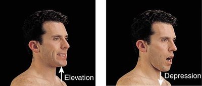
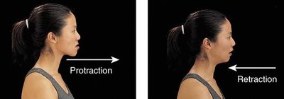
Mandible: Elevation (upward), depression (downward), protraction (forward), retraction (backward).
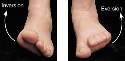
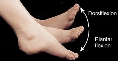
Hand and Foot: Inversion, eversion, dorsiflexion, plantarflexion, pronation, supination.
Summary Table: Types of Synovial Joints
Joint Type | Structure | Movement | Examples |
|---|---|---|---|
Ball-and-Socket | Ball fits into cuplike depression | Multiaxial (flexion, extension, abduction, adduction, rotation) | Shoulder, hip |
Hinge | Convex fits into concave | Uniaxial (flexion, extension) | Knee, elbow |
Pivot | Rounded bone with ring | Monoaxial (rotation) | Radioulnar, atlanto-axial |
Planar | Flat or slightly curved | Gliding | Intercarpal, intertarsal |
Condyloid | Oval projection into oval depression | Biaxial (flexion/extension, abduction/adduction) | Wrist, metacarpophalangeal |
Saddle | Saddle-shaped bone | Biaxial (circumduction, opposition) | Thumb |
Additional info: The notes above expand on brief points with academic context, definitions, and examples to ensure completeness and clarity for exam preparation.
