 Indietro
Articulations: Structure and Function of Joints
Guida di studio - Note intelligenti
 Appunti personalizzati basati sui tuoi materiali, ampliati con definizioni chiave, esempi e contesto.
Articulations (Joints)
Introduction to Joints
Joints, or articulations, are sites where two or more bones meet. They are essential for providing mobility to the skeleton while maintaining its integrity. Joints are classified both structurally and functionally, based on the materials binding the bones and the degree of movement allowed.
Structural classification: Fibrous, cartilaginous, and synovial joints
Functional classification: Synarthroses (immovable), amphiarthroses (slightly movable), diarthroses (freely movable)
Major Synovial Joints of the Body
Shoulder (Glenohumeral) Joint
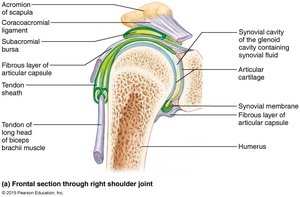
The shoulder joint is the most freely moving joint in the body, classified as a ball-and-socket synovial joint. Its structure allows for a wide range of motion but sacrifices stability, making it prone to dislocation.
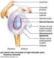
Articular surfaces: Large, hemispherical head of the humerus fits into the shallow glenoid cavity of the scapula.
Glenoid labrum: A fibrocartilaginous rim that deepens the cavity but still only holds about one-third of the humeral head.
Reinforcing ligaments: Coracohumeral ligament (supports upper limb), three glenohumeral ligaments (anterior capsule support).
Muscle tendons: The tendon of the long head of the biceps brachii acts as a 'superstabilizer.' Four rotator cuff muscles (subscapularis, supraspinatus, infraspinatus, teres minor) encircle the joint, providing most of its stability.
Clinical note: Dislocations are common, especially with lateral rotation and abduction, or from blows to the shoulder.
Elbow Joint
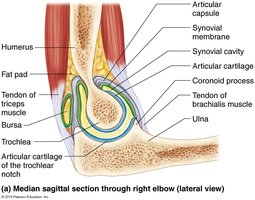
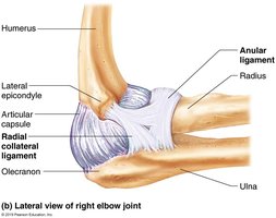
The elbow is a hinge joint formed primarily by the articulation between the trochlear notch of the ulna and the trochlea of the humerus. It allows for flexion and extension only.
Ligaments: Anular ligament (surrounds head of radius), ulnar collateral ligament, and radial collateral ligament (restrict side-to-side movement).
Articular capsule: Encloses the joint and is reinforced by surrounding muscles and ligaments.
Hip (Coxal) Joint
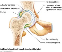
The hip joint is a ball-and-socket synovial joint, providing a good range of motion but with greater stability than the shoulder due to its deep socket.
Articular surfaces: Spherical head of the femur fits into the deep acetabulum of the hip bone.
Acetabular labrum: A rim of fibrocartilage that deepens the socket and enhances stability.
Reinforcing ligaments: Iliofemoral, pubofemoral, ischiofemoral ligaments, and the ligament of the head of the femur (ligamentum teres, which contains an artery for the femoral head).
Stability: Primarily from the deep socket and strong ligaments.
Knee Joint
The knee is the largest and most complex joint in the body, consisting of three joints within a single cavity: the femoropatellar joint and the lateral and medial tibiofemoral joints. It allows for flexion, extension, and some rotation when flexed.
Articular surfaces: Femoral condyles articulate with the tibial menisci.
Ligaments: Capsular, extracapsular, and intracapsular ligaments stabilize the joint. Key ligaments include the anterior and posterior cruciate ligaments (ACL and PCL), fibular and tibial collateral ligaments, oblique and arcuate popliteal ligaments.
Menisci: Medial and lateral menisci act as shock absorbers and improve the fit between femur and tibia.
Bursae: At least 12 bursae reduce friction around the knee.
Clinical note: The knee is vulnerable to horizontal blows, especially laterally, which can injure the 'three C's': collateral ligaments, cruciate ligaments, and cartilages (menisci).
The Pelvic Girdle
Structure and Function
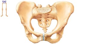
The pelvic girdle consists of two hip bones (coxal bones) and the sacrum. It attaches the lower limbs to the axial skeleton, transmits the weight of the upper body to the lower limbs, and supports pelvic organs. The pelvis is less mobile but more stable than the shoulder girdle.
Hip bone: Formed by the fusion of three bones: ilium, ischium, and pubis.
Acetabulum: Deep socket formed at the point of fusion, receives the head of the femur.
Pelvic Structure and Childbearing
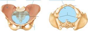
The pelvis is adapted for childbearing in females, being wider, shallower, lighter, and rounder than in males. The pelvic brim (inlet) and outlet are important landmarks for childbirth.
False pelvis: Superior to the pelvic brim.
True pelvis: Inferior to the pelvic brim; defines the birth canal.
Pelvic outlet: Inferior margin of the true pelvis.
Comparison of Male and Female Pelves
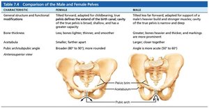
The male and female pelves differ in several anatomical features, reflecting their functional adaptations.
Characteristic | Female | Male |
|---|---|---|
General structure | Tilted forward, adapted for childbearing; broad, shallow, greater capacity | Tilted less far forward, adapted for support of heavier build; narrow, deep, less capacity |
Bones | Lighter, thinner, smoother | Heavier, thicker, markings more prominent |
Acetabula | Smaller, farther apart | Larger, closer together |
Pubic arch/angle | Broader (80–90°), more rounded | More acute (50–60°) |
Sacrum | Wider, shorter, less curved | Narrow, longer, more curved |
Coccyx | More movable, projects inferiorly | Less movable, projects anteriorly |
Pelvic inlet (brim) | Wider, oval from side to side | Narrow, basically heart-shaped |
Pelvic outlet | Wider, ischial spines farther apart | Narrower, ischial spines closer together |
Movements at Synovial Joints
Types of Movements
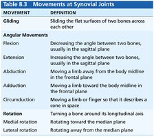
Synovial joints allow a variety of movements, classified as gliding, angular, and rotational movements. Each movement is defined by the change in angle or position of the bones involved.
Movement | Definition |
|---|---|
Gliding | Sliding the flat surfaces of two bones across each other |
Flexion | Decreasing the angle between two bones, usually in the sagittal plane |
Extension | Increasing the angle between two bones, usually in the sagittal plane |
Abduction | Moving a limb away from the body midline in the frontal plane |
Adduction | Moving a limb toward the body midline in the frontal plane |
Circumduction | Moving a limb or finger so that it describes a cone in space |
Rotation | Turning a bone around its longitudinal axis |
Medial rotation | Rotating toward the median plane |
Lateral rotation | Rotating away from the median plane |
Special Movements
Supination and pronation: Rotation of the radius and ulna (forearm).
Dorsiflexion and plantar flexion: Movements of the foot at the ankle.
Inversion and eversion: Movements of the sole of the foot medially or laterally.
Protraction and retraction: Anterior or posterior movement in a transverse plane (e.g., mandible).
Elevation and depression: Lifting or lowering a body part (e.g., shoulders, jaw).
Opposition: Movement of the thumb to touch the tips of other fingers.
Types of Synovial Joints
There are six types of synovial joints, classified by the shape of their articular surfaces and the movements they allow:
Plane (e.g., intercarpal joints)
Hinge (e.g., elbow joint)
Pivot (e.g., proximal radioulnar joint)
Condylar (e.g., metacarpophalangeal joints)
Saddle (e.g., carpometacarpal joint of the thumb)
Ball-and-socket (e.g., shoulder and hip joints)
Clinical Correlations
Shoulder dislocation: Due to the shallow glenoid cavity and high mobility, the shoulder is prone to dislocation, especially with lateral rotation and abduction.
Knee injuries: Lateral blows to the knee can damage the tibial collateral ligament, medial meniscus, and anterior cruciate ligament (the 'unhappy triad').
Arthroplasty: Surgical replacement of a joint.
Arthroscopy: Minimally invasive procedure using a camera to diagnose or repair joint structures.
