 Indietro
neuro 16
Guida di studio - Note intelligenti
 Appunti personalizzati basati sui tuoi materiali, ampliati con definizioni chiave, esempi e contesto.
Autonomic Nervous System
Anatomical and Cellular Differences
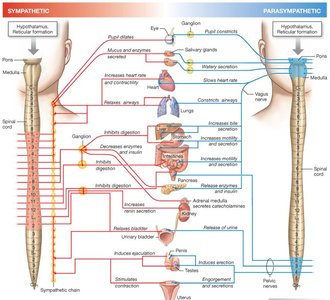
The autonomic nervous system (ANS) regulates involuntary physiological functions and is divided into the sympathetic and parasympathetic branches. Each branch has distinct anatomical origins and structural features.
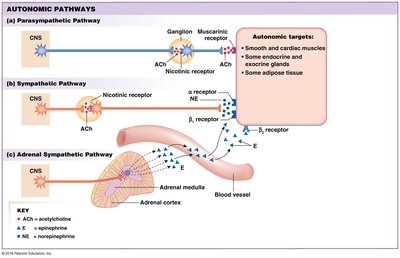
Sympathetic Division: Preganglionic neurons originate in the thoracic and lumbar regions of the spinal cord. Ganglia are located near the spinal cord (paravertebral).
Parasympathetic Division: Preganglionic neurons originate in the brainstem and sacral region. Ganglia are located on or near the target tissue.
Sympathetic pathways typically have short preganglionic and long postganglionic fibers, while parasympathetic pathways have long preganglionic and short postganglionic fibers.
Neuroeffector Junction
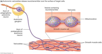
The neuroeffector junction is the site where autonomic neurons communicate with their target tissues.
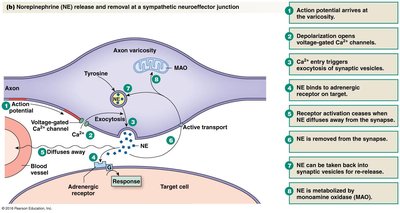
Autonomic neurotransmitters are released from varicosities along the axon, allowing for diffuse signaling.
Receptors are not clustered under varicosities; neurotransmitter diffuses to receptors on smooth muscle, cardiac muscle, glands, etc.
Neurotransmitters
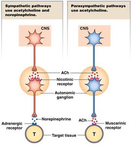
The ANS uses several neurotransmitters, primarily acetylcholine (ACh) and norepinephrine (NE).
Sympathetic Pathways: Preganglionic neurons release ACh; postganglionic neurons usually release NE.
Parasympathetic Pathways: Both pre- and postganglionic neurons release ACh.
Some sympathetic neurons are cholinergic (release ACh), and some autonomic neurons use other neurotransmitters (e.g., ATP, NO).
Adrenal Medulla and Neuroendocrine Signaling
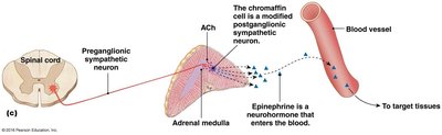
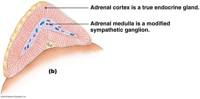
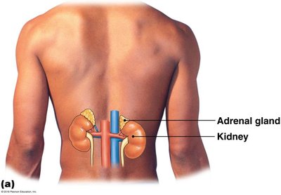
The adrenal medulla is a specialized neuroendocrine structure associated with the sympathetic nervous system.
It acts as a modified sympathetic ganglion, containing chromaffin cells (modified postganglionic neurons).
Upon stimulation, it releases catecholamines (mainly epinephrine) into the bloodstream, affecting distant target tissues.
Synthesis and Release of Neurotransmitters
Norepinephrine: Synthesized from tyrosine in axon varicosities; release is Ca2+-mediated and terminated by reuptake or enzymatic degradation (monoamine oxidase).
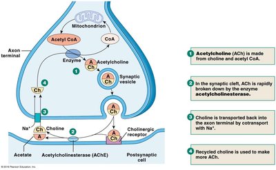
Acetylcholine: Synthesized from choline and acetyl CoA by choline acetyltransferase; broken down by acetylcholinesterase in the synaptic cleft.
Autonomic Receptors and Signaling Pathways
Autonomic receptors are classified as adrenergic (sympathetic) or cholinergic (parasympathetic), with multiple subtypes.
Adrenergic Receptors: All are G-protein coupled (metabotropic); main types are alpha (α) and beta (β), each with subtypes.
Cholinergic Receptors: Nicotinic (ionotropic) and muscarinic (metabotropic, G-protein coupled).
Muscarinic receptors (M1–M5) are found on parasympathetic target tissues; M2 and M3 are most common.
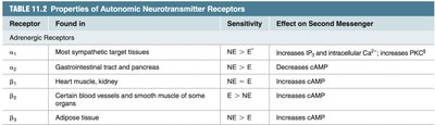

G-Protein Coupled Receptor Signaling
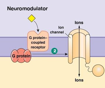
G-protein coupled receptors (GPCRs) mediate cellular responses via two main pathways:
Ion Channel Modulation: GPCRs can open or close ion channels, affecting membrane potential.
Enzyme Activation: GPCRs activate membrane-bound enzymes, leading to second messenger cascades.
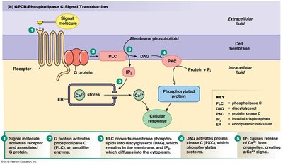
Phospholipase C Signal Transduction Pathway
Activation of phospholipase C (PLC) increases intracellular Ca2+ and activates protein kinase C (PKC), leading to cellular responses.
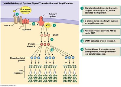
Adenylyl Cyclase Signal Transduction Pathway
Activation of adenylyl cyclase increases cyclic AMP (cAMP), which activates protein kinase A (PKA) and leads to protein phosphorylation and cellular responses.
Summary of Autonomic Pathways
Autonomic pathways consist of a preganglionic and postganglionic neuron in series, except for the adrenal medulla.
Sympathetic division is most active during stress; parasympathetic during rest-and-digest.
Sympathetic postganglionic neurons release NE; parasympathetic postganglionic neurons release ACh.
Muscle Types
Introduction to Muscle Types
Muscle tissue is specialized for generating force and movement in response to physiological stimuli.
Functions include body movement, posture, respiration, heat production, communication, constriction of organs/vessels, and heartbeat.
All muscle types rely on the conversion of chemical energy (ATP) to mechanical energy.
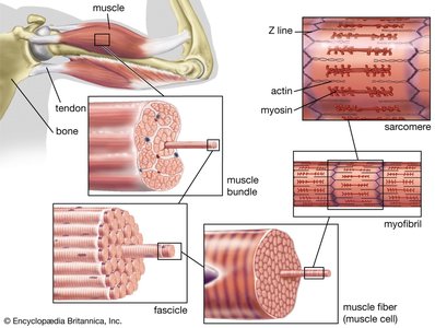
Skeletal Muscle
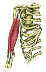
Primarily voluntary, controlled by somatic motor neurons.
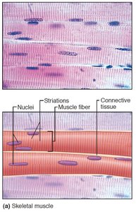
Striated and multinucleated.
Responsible for movement, posture, and heat production.
Cardiac Muscle
Primarily involuntary; exhibits spontaneous electrical activity.
Striated and uninucleated.
Can be modulated by the autonomic nervous system and hormones.
Smooth Muscle
Primarily involuntary; controlled by autonomic nervous system, hormones, paracrines, or autocrines.
Non-striated and uninucleated.
Provides mechanical control of digestive, urinary, reproductive tracts, blood vessels, and airways.
Summary Table: Muscle Types
Muscle Type | Control | Striations | Nuclei | Main Function |
|---|---|---|---|---|
Skeletal | Voluntary | Striated | Multinucleated | Movement, posture |
Cardiac | Involuntary | Striated | Uninucleated | Heartbeat |
Smooth | Involuntary | Non-striated | Uninucleated | Organ/vessel constriction |
Example: Muscle Function
Skeletal muscle contracts to move bones and maintain posture.
Cardiac muscle contracts rhythmically to pump blood.
Smooth muscle contracts to regulate diameter of blood vessels and propel contents through organs.
Additional info:
ATP hydrolysis is the universal mechanism for force generation in all muscle types.
Muscle contraction is regulated by calcium ions and specific signaling pathways.
