 Indietro
Autonomic Nervous System Study Guide: Step-by-Step Guidance
Guida di studio - Note intelligenti
 Appunti personalizzati basati sui tuoi materiali, ampliati con definizioni chiave, esempi e contesto.
Q1. Compare the organization of the autonomic nervous system with that of the somatic nervous system.
Background
Topic: Nervous System Organization
This question tests your understanding of the structural and functional differences between the somatic and autonomic nervous systems, including their effectors and control mechanisms.
Key Terms:
Somatic Nervous System: Controls voluntary movements via skeletal muscle.
Autonomic Nervous System: Controls involuntary functions via smooth muscle, cardiac muscle, glands, and adipose tissue.
Effector: The target tissue or organ that responds to neural stimulation.
Neuron: A nerve cell; in this context, refers to lower motor neurons and ganglionic neurons.
Step-by-Step Guidance
Identify the types of tissues controlled by each system (somatic vs. autonomic).
Determine where the neurons that exert direct control over effectors are located for each system.
Summarize the main effectors for the somatic system (skeletal muscle) and for the autonomic system (smooth muscle, cardiac muscle, glands, adipose tissue).
Describe the location of lower motor neurons in the CNS for the somatic system and ganglionic neurons in autonomic ganglia for the autonomic system.
Set up a comparison table to organize these differences.
Try solving on your own before revealing the answer!
Final Answer:
Somatic Nervous System controls skeletal muscle via lower motor neurons in the CNS (brain stem & spinal cord). Autonomic Nervous System controls smooth muscle, cardiac muscle, glands, and adipose tissue via ganglionic neurons in autonomic ganglia outside the CNS.
The neurons exerting direct control are lower motor neurons (somatic) and ganglionic neurons (autonomic).
Q2. What part of the brain contains the integrative centers for autonomic activity?
Background
Topic: Central Nervous System Control
This question is about the location of higher-level control centers for autonomic functions in the brain.
Key Terms:
Integrative Centers: Areas in the brain that coordinate autonomic functions.
Hypothalamus: A region of the brain involved in autonomic regulation.
Step-by-Step Guidance
Recall which brain region is responsible for integrating autonomic functions.
Consider the role of the hypothalamus and other brain stem centers.
Think about how these centers interact with the rest of the nervous system.
Try solving on your own before revealing the answer!
Final Answer:
The hypothalamus contains the integrative centers for autonomic activity, with additional centers in the brain stem/medulla oblongata.
Q3. Where in the CNS are the cell bodies of preganglionic neurons found?
Background
Topic: Neuron Anatomy
This question tests your knowledge of the anatomical location of preganglionic neuron cell bodies in the central nervous system.
Key Terms:
Preganglionic Neuron: The first neuron in the autonomic pathway.
Visceral Motor Nuclei: Clusters of neuron cell bodies in the CNS.
Lateral Gray Horns: Specific region of the spinal cord.
Step-by-Step Guidance
Identify the regions of the CNS where autonomic preganglionic neurons originate.
Recall the difference between somatic and autonomic neuron locations.
Consider both the brain stem and spinal cord regions.
Try solving on your own before revealing the answer!
Final Answer:
Preganglionic neuron cell bodies are found in visceral motor (autonomic) nuclei of the brain stem and in the lateral gray horns of the spinal cord.
Q4. Where are the cell bodies of ganglionic neurons found?
Background
Topic: Peripheral Nervous System Anatomy
This question is about the location of ganglionic neuron cell bodies in the autonomic nervous system.
Key Terms:
Ganglionic Neuron: The second neuron in the autonomic pathway.
Autonomic Ganglia: Clusters of neuron cell bodies outside the CNS.
Step-by-Step Guidance
Recall the definition of ganglionic neurons and their role in the autonomic pathway.
Identify where autonomic ganglia are located relative to the CNS.
Think about the difference between central and peripheral nervous system structures.
Try solving on your own before revealing the answer!
Final Answer:
Ganglionic neuron cell bodies are found in autonomic ganglia in the peripheral nervous system (outside the CNS).
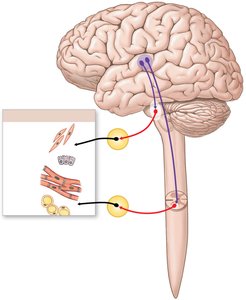
Q5. On the figure above, label the following regions/structures: four visceral effectors, motor nuclei in hypothalamus, autonomic nuclei in brain stem, autonomic nuclei in spinal cord, preganglionic neurons, autonomic ganglia, ganglionic neurons.
Background
Topic: Autonomic Pathways and Anatomy
This question tests your ability to identify and label key anatomical structures involved in autonomic nervous system pathways.
Key Terms:
Visceral Effectors: Target tissues/organs (smooth muscle, glands, cardiac muscle, adipose tissue).
Motor Nuclei: CNS clusters controlling autonomic output.
Preganglionic/Ganglionic Neurons: Neurons in the autonomic pathway.
Autonomic Ganglia: Peripheral clusters of neuron cell bodies.
Step-by-Step Guidance
Examine the diagram and identify the brain regions (hypothalamus, brain stem, spinal cord).
Locate the autonomic nuclei in the brain stem and spinal cord.
Trace the pathway from motor nuclei to preganglionic neurons, then to autonomic ganglia and ganglionic neurons.
Identify the four visceral effectors in the diagram (smooth muscle, glands, cardiac muscle, adipose tissue).
Label each structure according to its role in the pathway.
Try labeling on your own before revealing the answer!
Final Answer:
Four visceral effectors: smooth muscle, glands, cardiac muscle, adipose tissue
Motor nuclei in hypothalamus
Autonomic nuclei in brain stem
Autonomic nuclei in spinal cord
Preganglionic neurons (red fibers)
Autonomic ganglia (yellow circles)
Ganglionic neurons (black fibers)
Q6. Summarize the general effects of the sympathetic and parasympathetic divisions of the autonomic nervous system.
Background
Topic: Autonomic Nervous System Function
This question is about the physiological effects of the two main divisions of the autonomic nervous system.
Key Terms:
Sympathetic Division: "Fight or flight" responses.
Parasympathetic Division: "Rest and digest" responses.
Step-by-Step Guidance
Recall the main functions of the sympathetic division (increased alertness, heart rate, blood pressure, etc.).
Recall the main functions of the parasympathetic division (conserves energy, slows heart rate, stimulates digestion, etc.).
Summarize these effects in your own words.
Try summarizing on your own before revealing the answer!
Final Answer:
Sympathetic: "Fight or flight" — increases alertness, metabolic rate, heart rate and blood pressure; dilates airways and pupils; mobilizes energy reserves; shunts blood to skeletal muscles; slows digestion and urination.
Parasympathetic: "Rest and digest" — conserves energy and promotes sedentary activities: slows heart rate, constricts pupils, stimulates digestion, salivation and nutrient absorption, and promotes urination and defecation.
Q7. Define cholinergic. Which neurons in the sympathetic division are cholinergic?
Background
Topic: Neurotransmitter Release
This question is about the definition of cholinergic neurons and their role in the sympathetic division.
Key Terms:
Cholinergic: Neurons that release acetylcholine (ACh).
Preganglionic Neurons: First neuron in the autonomic pathway.
Step-by-Step Guidance
Define what it means for a neuron to be cholinergic.
Identify which neurons in the sympathetic division release ACh.
Recall the role of preganglionic neurons and any exceptions among ganglionic neurons.
Try answering on your own before revealing the answer!
Final Answer:
A cholinergic neuron releases acetylcholine (ACh) as its neurotransmitter. All preganglionic neurons (plus a few ganglionic neurons, e.g., those to sweat glands and some blood vessels in skeletal muscle) in the sympathetic division are cholinergic.
Q8. What effect do cholinergic neurons have on ganglionic neurons? Do the ganglionic neurons undergo an EPSP or IPSP?
Background
Topic: Synaptic Transmission
This question tests your understanding of the effect of acetylcholine on ganglionic neurons in the autonomic nervous system.
Key Terms:
EPSP: Excitatory postsynaptic potential.
IPSP: Inhibitory postsynaptic potential.
Nicotinic Receptors: ACh receptors on ganglionic neurons.
Step-by-Step Guidance
Recall the neurotransmitter released by preganglionic neurons (ACh).
Identify the type of receptor on ganglionic neurons (nicotinic).
Determine whether the effect is excitatory or inhibitory.
Try answering on your own before revealing the answer!
Final Answer:
ACh binds nicotinic receptors on the ganglionic neurons and is always excitatory, so the ganglionic neurons undergo an EPSP.
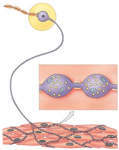
Q9. On the figure, label the following: smooth muscle fibers, varicosities, vesicles containing NE, ganglion, ganglionic neuron, postganglionic fiber, preganglionic fiber.
Background
Topic: Sympathetic Neurotransmitter Release
This question tests your ability to identify and label structures involved in neurotransmitter release in the sympathetic division.
Key Terms:
Varicosity: Swelling along a postganglionic axon.
NE: Norepinephrine, a neurotransmitter.
Ganglion: Cluster of neuron cell bodies.
Postganglionic Fiber: Axon of ganglionic neuron.
Step-by-Step Guidance
Examine the diagram and identify the ganglion and ganglionic neuron.
Trace the preganglionic and postganglionic fibers.
Locate the varicosities and vesicles containing NE along the postganglionic fiber.
Identify the smooth muscle fibers as the target tissue.
Label each structure according to its role in neurotransmitter release.
Try labeling on your own before revealing the answer!
Final Answer:
Smooth muscle fibers (bottom tissue)
Varicosities (swelling along postganglionic fiber)
Vesicles containing NE (inside varicosities)
Ganglion (yellow circle)
Ganglionic neuron (cell body in ganglion)
Postganglionic fiber (axon extending from ganglion)
Preganglionic fiber (axon leading to ganglion)
Q10. How is a varicosity different from a regular axon terminal?
Background
Topic: Neurotransmitter Release Mechanisms
This question is about the structural differences between varicosities and typical axon terminals in neurotransmitter release.
Key Terms:
Varicosity: Swelling along an axon that releases neurotransmitter.
Axon Terminal: The end of a neuron where neurotransmitter is released.
Step-by-Step Guidance
Recall the structure of a typical axon terminal (single knob at the end).
Compare this to the structure of varicosities (multiple swellings along the axon).
Consider how neurotransmitter release differs between the two.
Try answering on your own before revealing the answer!
Final Answer:
A varicosity is a swelling along a branched postganglionic axon, not a single knob at its end. Each axon has many varicosities strung like beads, releasing NE en passant onto a broad area of smooth muscle; there is no specialized, tight synapse, so NE diffuses to many effector cells.
