 Indietro
Axial and Appendicular Skeleton: Structure and Function
Guida di studio - Note intelligenti
 Appunti personalizzati basati sui tuoi materiali, ampliati con definizioni chiave, esempi e contesto.
The Human Skeleton: Overview
Axial and Appendicular Skeleton
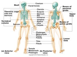
The human skeleton is divided into two main parts: the axial skeleton and the appendicular skeleton. The axial skeleton forms the central axis of the body and includes the skull, vertebral column, and thoracic cage. The appendicular skeleton consists of the bones of the limbs and the girdles that attach them to the axial skeleton.
Axial skeleton: Skull, vertebral column, rib cage (sternum and ribs)
Appendicular skeleton: Upper and lower limbs, pectoral (shoulder) girdle, pelvic (hip) girdle
Total bones in adult human: 206
Skull: Structure and Key Bones
Major Bones and Regions of the Skull
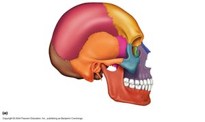
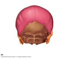
The skull is a complex structure composed of cranial and facial bones. It protects the brain and supports the structures of the face.
Cranial bones: Enclose the brain (e.g., frontal, parietal, temporal, occipital, sphenoid, ethmoid)
Facial bones: Form the framework of the face (e.g., maxilla, mandible, zygomatic, nasal)
Sutures: Immovable joints connecting skull bones
Temporal Bone
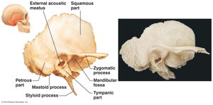
The temporal bone is an irregularly shaped bone on the side of the skull. It is the site of attachment for neck and tongue muscles and houses the middle and inner ear cavities, which are located in the petrous part—the hardest bone in the body.
Petrous part: Contains organs for hearing and balance
Processes: Mastoid, styloid, zygomatic
Sphenoid Bone
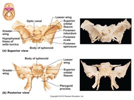
The sphenoid bone is a complex, bat-shaped bone that articulates with all other cranial bones. It forms part of the floor of the cranial cavity and contains important foramina for nerves and blood vessels.
Greater and lesser wings
Body of sphenoid
Optic canal, sella turcica
Ethmoid Bone
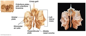
The ethmoid bone is a cube-shaped bone located deep between the orbits and nasal cavities. The crista galli projects superiorly to attach to the dura mater of the brain.
Cribriform plate: Contains foramina for olfactory nerves
Perpendicular plate: Forms part of the nasal septum
Middle nasal concha: Increases surface area in the nasal cavity
Sutural Bones
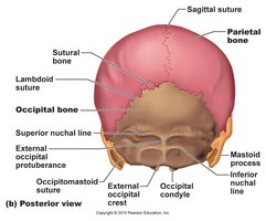
Sutural bones are tiny, irregular bones that can occur within cranial sutures. They result from additional ossification centers during fetal development.
Hyoid Bone
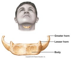
The hyoid bone is unique because it does not articulate directly with any other bone. It is anchored by ligaments and serves as a movable base for the tongue and an attachment site for muscles involved in swallowing and speech. It is horseshoe-shaped and can be fractured by strangulation.
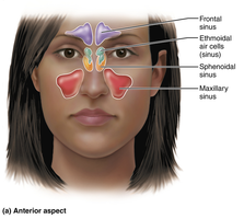
Paranasal Sinuses
Paranasal sinuses are air-filled spaces in the skull that lighten the skull, moisten and warm incoming air, and contribute to voice resonance.
Frontal, ethmoidal, sphenoidal, and maxillary sinuses
Vertebral Column
Structure and Regions
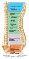
The vertebral column (spine) extends from the skull to the pelvis. It transmits the weight of the trunk to the lower limbs, protects the spinal cord, and provides attachment points for ribs and muscles. It consists of 33 bones, of which 24 remain separate and 9 fuse to form the sacrum and coccyx.
Cervical region: 7 vertebrae (C1–C7)
Thoracic region: 12 vertebrae (T1–T12)
Lumbar region: 5 vertebrae (L1–L5)
Sacrum: 5 fused vertebrae
Coccyx: 3–5 fused vertebrae
Ligaments and Intervertebral Discs
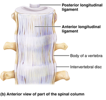
Ligaments support the vertebral column and keep it upright. The major supporting ligaments are the anterior and posterior longitudinal ligaments. Intervertebral discs are cushion-like pads between vertebrae that act as shock absorbers.
Anterior longitudinal ligament: Broad, strong, prevents hyperextension
Posterior longitudinal ligament: Narrow, weaker, prevents hyperflexion
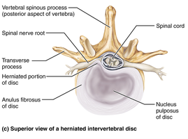
Intervertebral disc: Consists of anulus fibrosus (outer ring) and nucleus pulposus (inner gel-like center)
General Structure of Vertebrae
All vertebrae share common features:
Body: Anterior, weight-bearing region
Vertebral arch: Composed of pedicles and laminae
Vertebral foramen: Encloses the spinal cord
Intervertebral foramina: Lateral openings for spinal nerves
Sacrum and Coccyx
The sacrum is formed by the fusion of five vertebrae and serves as a central support for the skeleton, housing nerves such as the pelvic nerves. The coccyx (tailbone) is formed from three to five fused vertebrae and provides support while sitting and serves as an anchor for pelvic floor and anal muscles.
Thoracic Cage: Sternum and Ribs
Thoracic Cage Structure and Function
The thoracic cage consists of the thoracic vertebrae, ribs, sternum, and costal cartilages. It protects vital organs, supports the shoulders and upper limbs, and provides attachment sites for muscles.
Sternum: Breastbone, composed of manubrium, body, and xiphoid process
Ribs: 12 pairs, each with a shaft, head, neck, and tubercle
Appendicular Skeleton
Pectoral Girdle
The pectoral girdle attaches the upper limbs to the trunk and consists of the clavicle and scapula. These bones are light and allow for a wide range of motion.
Pelvic Girdle
The pelvic girdle attaches the lower limbs to the trunk and is formed by two hip bones (coxal bones) and the sacrum. It transmits the weight of the upper body to the lower limbs and supports pelvic organs. The pelvic girdle is less mobile but more stable than the shoulder joint.
Bones of the Upper Limb
Arm: Humerus (longest bone in upper limb)
Forearm: Radius and ulna (parallel long bones)
Hand: 8 carpals (wrist), 5 metacarpals (palm), 14 phalanges (fingers)
Bones of the Lower Limb
Thigh: Femur (longest bone in body)
Leg: Tibia (medial, weight-bearing) and fibula (lateral, non-weight-bearing)
Foot: Tarsals, metatarsals, phalanges
Pelvis: True vs. False Pelvis and Sex Differences
The true pelvis is the region below the pelvic brim and is involved in childbirth, while the false pelvis is above the brim and supports abdominal organs. Male and female pelves differ structurally to accommodate childbirth in females.
Summary Table: Major Bones to Know
Region | Major Bones |
|---|---|
Skull | Frontal, parietal, temporal, occipital, sphenoid, ethmoid, maxilla, mandible, zygomatic, nasal |
Vertebral Column | Cervical, thoracic, lumbar vertebrae, sacrum, coccyx |
Thoracic Cage | Sternum, ribs |
Pectoral Girdle | Clavicle, scapula |
Upper Limb | Humerus, radius, ulna, carpals, metacarpals, phalanges |
Pelvic Girdle | Hip bones (coxal bones), sacrum |
Lower Limb | Femur, tibia, fibula, patella, tarsals, metatarsals, phalanges |
Key Definitions
Axial skeleton: Central skeleton including skull, vertebral column, and thoracic cage
Appendicular skeleton: Limbs and girdles attaching them to the axial skeleton
Suture: Immovable joint between skull bones
Foramen: Opening in bone for nerves and blood vessels
Process: Projection or outgrowth of bone
Practice Questions
What is not part of the axial skeleton? (Answer: Clavicle)
What is the correct statement about the humerus? (Answer: Only bone in the ‘arm’)
What is true of the Pectoral Girdle? (Answer: Contains light bones that are easy to move)
The number of bones in the human body is: (Answer: 206)
What is the false statement about the skull? (Answer: Ethmoid bone is said to be bat shaped)
Strangulation may be expected to result in fracturing of the: (Answer: Hyoid bone)
