 Indietro
Axial and Appendicular Skeleton: Structure and Function
Guida di studio - Note intelligenti
 Appunti personalizzati basati sui tuoi materiali, ampliati con definizioni chiave, esempi e contesto.
Skeletal System Overview
Axial and Appendicular Skeleton
The human skeleton is divided into two main regions: the axial skeleton and the appendicular skeleton. The axial skeleton forms the central axis of the body, while the appendicular skeleton consists of the limbs and girdles that attach them to the axial skeleton.
Axial Skeleton: Includes the skull, vertebral column, and thoracic cage.
Appendicular Skeleton: Includes the pectoral and pelvic girdles, and the upper and lower limbs.
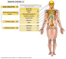
Axial Skeleton
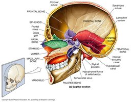
Skull Structure
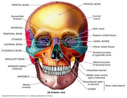
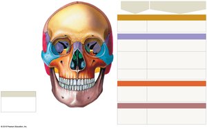
The skull is composed of cranial bones, which enclose the cranial cavity, and facial bones, which surround and protect the entrances to the respiratory and digestive tracts. Associated bones include the auditory ossicles and the hyoid bone.
Cranial Bones: Occipital, parietal, frontal, temporal, sphenoid, and ethmoid.
Facial Bones: Maxillary, palatine, nasal, vomer, zygomatic, lacrimal, inferior nasal concha, and mandible.
Associated Bones: Auditory ossicles (malleus, incus, stapes) and hyoid bone.
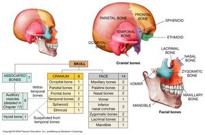
Skull Sutures and Landmarks
Sutures are immovable joints between skull bones. Major sutures include the coronal, sagittal, lambdoidal, and squamous sutures. These landmarks are important for anatomical orientation and clinical reference.
Coronal Suture: Between frontal and parietal bones.
Sagittal Suture: Between the two parietal bones.
Lambdoidal Suture: Between parietal and occipital bones.
Squamous Suture: Between parietal and temporal bones.
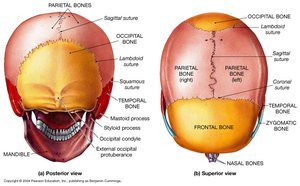
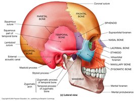
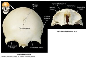
Frontal Bone
The frontal bone forms the forehead and the superior part of the orbital cavity. It contains the frontal sinuses and is bordered by the coronal suture.
Frontal Squama: Flat part forming the forehead.
Supraorbital Margin: Superior border of the eye socket.
Frontal Sinus: Air-filled cavity within the bone.
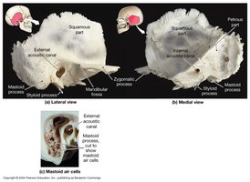
Temporal Bone
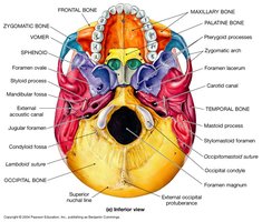
The temporal bone forms part of the lateral and base of the skull. It houses structures of the ear and provides attachment points for muscles.
Mastoid Process: Contains air cells, attachment for neck muscles.
External Acoustic Canal: Passage for sound waves.
Zygomatic Process: Articulates with zygomatic bone.
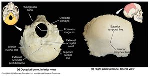
Occipital Bone
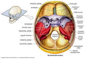
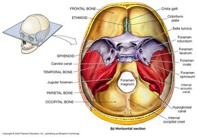
The occipital bone forms the posterior and base of the skull. It contains the foramen magnum, through which the spinal cord passes.
Foramen Magnum: Large opening for spinal cord.
Occipital Condyles: Articulate with the first cervical vertebra.
External Occipital Protuberance: Prominent midline bump.
Sphenoid and Ethmoid Bones
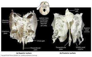
The sphenoid bone forms part of the cranial floor and contains the sella turcica, which houses the pituitary gland. The ethmoid bone forms part of the nasal cavity and the medial wall of the orbit.
Sphenoid: Contains optic canal, sella turcica, and pterygoid processes.
Ethmoid: Contains crista galli, cribriform plate, and perpendicular plate.
Facial Bones
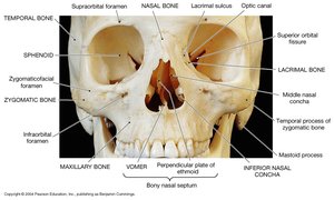
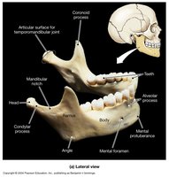
Facial bones provide structure for the face and protect the entrances to the respiratory and digestive systems. The mandible is the only movable bone of the skull.
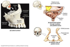
Maxillae: Form upper jaw and most of hard palate.
Mandible: Lower jaw, only movable joint in skull.
Palatine Bones: Form posterior hard palate and floor of nasal cavity.
Nasal Bones: Form bridge of nose.
Zygomatic Bones: Form cheekbones.
Lacrimal Bones: Smallest facial bones, part of medial orbit.
Vomer: Forms part of nasal septum.
Auditory Ossicles and Hyoid Bone
The auditory ossicles are three small bones in the middle ear (malleus, incus, stapes) that transmit sound vibrations. The hyoid bone is located in the neck and supports the tongue and larynx.
Auditory Ossicles: Malleus, incus, stapes.
Hyoid Bone: Does not articulate with other bones; supports tongue.
Vertebral Column
Structure and Regions
The vertebral column consists of 33 vertebrae, grouped into cervical, thoracic, lumbar, sacral, and coccygeal regions. It provides support and protection for the spinal cord and allows flexibility and movement.
Cervical: 7 vertebrae (C1–C7)
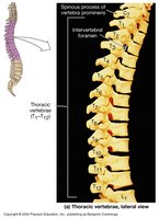
Thoracic: 12 vertebrae (T1–T12)
Lumbar: 5 vertebrae (L1–L5)
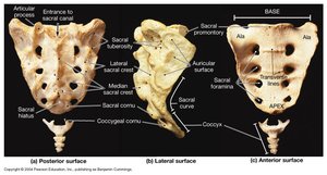
Sacrum: 5 fused vertebrae
Coccyx: 3–5 fused vertebrae
Spinal Curvatures
The vertebral column has four curvatures: cervical, thoracic, lumbar, and sacral. These curves help distribute mechanical stress during movement and standing.
Primary Curvatures: Thoracic and sacral, present at birth.
Secondary Curvatures: Cervical (develops when baby holds up head) and lumbar (develops when baby walks).
Vertebral Anatomy
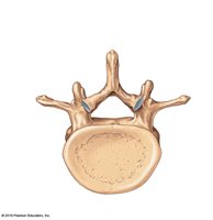
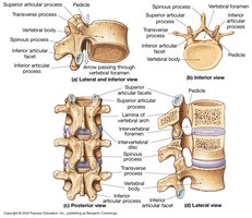
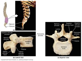
Each vertebra consists of a body, vertebral arch, and several processes for muscle attachment and articulation.
Body: Main weight-bearing portion.
Vertebral Arch: Forms vertebral foramen for spinal cord.
Processes: Spinous, transverse, and articular processes.
Intervertebral Discs: Fibrocartilage pads between vertebrae.
Regional Vertebral Differences
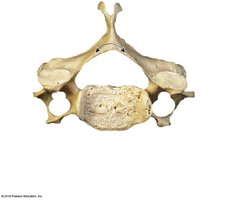
Vertebrae differ in shape and features depending on their region.
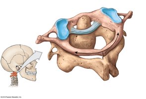
Cervical: Smallest, have transverse foramina, bifurcated spinous processes.
Thoracic: Larger, have facets for rib articulation, long spinous processes.
Lumbar: Largest, thick body, short spinous process.
Thoracic Cage
Rib Cage Structure
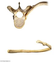
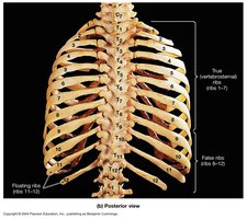
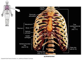
The thoracic cage consists of the ribs and sternum, protecting the heart and lungs and providing attachment points for muscles involved in respiration.
Ribs: 12 pairs; true ribs (1–7), false ribs (8–12), floating ribs (11–12).
Sternum: Manubrium, body, xiphoid process.
Appendicular Skeleton
Pectoral Girdle
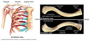
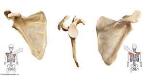
The pectoral girdle consists of the clavicle and scapula, connecting the upper limb to the trunk. It allows for a wide range of motion but is less stable than the pelvic girdle.
Clavicle: Articulates with sternum (medial) and scapula (lateral).
Scapula: Held in place by muscles; features include acromion and coracoid processes.
Upper Limb
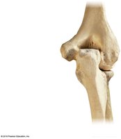
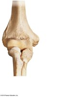
The upper limb includes the humerus, radius, ulna, carpal bones, metacarpals, and phalanges. The humerus articulates with the scapula at the glenohumeral joint.
Humerus: Features include greater and lesser tubercles, deltoid tuberosity, capitulum, trochlea, and olecranon fossa.
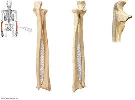
Radius: Lateral bone of forearm; radial tuberosity for muscle attachment.
Ulna: Medial bone; features include olecranon and coronoid processes.
Hand and Wrist
The hand consists of carpal bones (wrist), metacarpals (palm), and phalanges (fingers). The thumb (pollex) has two phalanges, while other fingers have three.
Carpal Bones: Two rows of short bones.
Metacarpals: Five bones forming the palm.
Phalanges: Proximal, middle, and distal segments.
Pelvic Girdle
The pelvic girdle consists of two os coxae bones, each formed by the ilium, ischium, and pubis. It supports the weight of the upper body and protects pelvic organs.
Ilium: Largest hip bone; features include iliac crest and sciatic notches.
Ischium: Contains ischial tuberosity.
Pubis: Forms pubic symphysis and ramus.
Acetabulum: Socket for femur head.
Male vs. Female Pelvis
The male and female pelvis differ in structure to accommodate childbirth in females.
Male: Iliac crests and acetabula close together, pubic arch less than 90°, curved sacrum and coccyx.
Female: Iliac crests and acetabula further apart, pubic arch greater than 90°, flatter sacrum and coccyx.
Lower Limb
The lower limb includes the femur, patella, tibia, fibula, tarsal bones, metatarsals, and phalanges. The femur is the longest and heaviest bone in the body.
Femur: Features include head, neck, trochanters, condyles, and linea aspera.
Patella: Sesamoid bone protecting the knee joint.
Tibia: Main weight-bearing bone of the leg.
Fibula: Lateral bone, not part of knee joint.
Ankles and Feet
The ankle contains seven tarsal bones. The foot has a similar pattern to the hand, with metatarsals and phalanges. The big toe (hallux) has two phalanges, while other toes have three.
Summary Table: Axial vs. Appendicular Skeleton
Region | Main Components | Function |
|---|---|---|
Axial Skeleton | Skull, vertebral column, thoracic cage | Protection, support, muscle attachment |
Appendicular Skeleton | Pectoral girdle, pelvic girdle, limbs | Movement, manipulation, support |
Key Equations
Bone count in adult human skeleton:
Vertebral column regions:
Additional info: Academic context and expanded explanations were added to ensure completeness and clarity for exam preparation.
