 Indietro
Axial Muscles: Structure, Function, and Innervation
Guida di studio - Note intelligenti
 Appunti personalizzati basati sui tuoi materiali, ampliati con definizioni chiave, esempi e contesto.
Axial Muscles
Introduction to Axial Muscles
Axial muscles are those that are located along the central axis of the body, including the head, neck, thorax, abdomen, and back. They play essential roles in movement, posture, respiration, and protection of internal organs.
Definition: Axial muscles are muscles that attach to the axial skeleton and are responsible for movements of the head, neck, trunk, and respiration.
Examples: Muscles of facial expression, mastication, pharyngeal muscles, neck muscles, respiratory muscles, abdominal muscles, and back muscles.
Muscle Actions
Muscles produce movement by contracting and pulling on bones or skin.
Flexion: Decreases the angle between two body parts (e.g., biceps brachii).
Extension: Increases the angle between two body parts (e.g., triceps brachii).
Abduction: Moves a limb away from the midline (e.g., deltoid).
Adduction: Moves a limb toward the midline (e.g., pectoralis major).
Rotation: Turns a body part around its axis (e.g., sternocleidomastoid).
Circular/Sphincter: Muscles that surround openings and control passage (e.g., orbicularis oris).
Muscle Fascicle Arrangements
The arrangement of muscle fibers (fascicles) determines the muscle's range of motion and power.
Parallel: Fascicles run parallel to the long axis of the muscle (e.g., sartorius).
Pennate: Fascicles attach obliquely to a central tendon (e.g., rectus femoris).
Circular: Fascicles arranged in concentric rings (e.g., orbicularis oris).
Muscles of Facial Expression
Overview
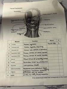
Muscles of facial expression are responsible for movements of the skin and soft tissues of the face, allowing for expressions such as smiling, frowning, and blinking. Most are innervated by the facial nerve (CN VII).
Orbicularis oculi: Closes eyelids, squinting (CN VII).
Orbicularis oris: Closes lips, purses lips (CN VII).
Zygomaticus major: Elevates corners of mouth (smiling) (CN VII).
Buccinator: Compresses cheek, holds food between teeth (CN VII).
Frontalis: Raises eyebrows, wrinkles forehead (CN VII).
Depressor anguli oris: Depresses corner of mouth (CN VII).
Muscle | Function | Nerve |
|---|---|---|
Orbicularis oculi | Closes eyelids, squinting | CN VII |
Orbicularis oris | Closes lips, purses lips | CN VII |
Zygomaticus major | Elevates corners of mouth | CN VII |
Buccinator | Compresses cheek | CN VII |
Frontalis | Raises eyebrows | CN VII |
Depressor anguli oris | Depresses corner of mouth | CN VII |
Muscles of Mastication
Overview
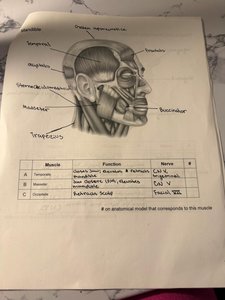
Muscles of mastication are responsible for movements of the jaw during chewing. They are innervated by the trigeminal nerve (CN V).
Temporalis: Elevates and retracts mandible (CN V).
Masseter: Elevates mandible, closes jaw (CN V).
Buccinator: Compresses cheek, assists in chewing (CN VII).
Occipitofrontalis: Moves scalp (CN VII).
Muscle | Function | Nerve |
|---|---|---|
Temporalis | Elevates & retracts mandible | CN V |
Masseter | Elevates mandible | CN V |
Buccinator | Compresses cheek | CN VII |
Occipitofrontalis | Moves scalp | CN VII |
Tongue and Pharyngeal Muscles
Overview
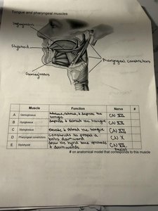
These muscles are involved in swallowing, speech, and movement of the tongue. They are innervated by various cranial nerves.
Genioglossus: Protrudes and depresses tongue (CN XII).
Styloglossus: Elevates and retracts tongue (CN XII).
Hyoglossus: Depresses and retracts tongue (CN XII).
Pharyngeal constrictors: Constrict pharynx during swallowing (CN X).
Muscle | Function | Nerve |
|---|---|---|
Genioglossus | Protrudes, depresses tongue | CN XII |
Styloglossus | Elevates, retracts tongue | CN XII |
Hyoglossus | Depresses, retracts tongue | CN XII |
Pharyngeal constrictors | Constrict pharynx | CN X |
Muscles of the Head and Neck
Overview
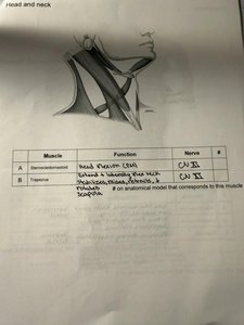
These muscles control movements of the head and neck, including flexion, extension, and rotation.
Sternocleidomastoid: Flexes and rotates the head (CN XI).
Trapezius: Extends neck, elevates scapula (CN XI).
Muscle | Function | Nerve |
|---|---|---|
Sternocleidomastoid | Head flexion, rotation | CN XI |
Trapezius | Extends neck, elevates scapula | CN XI |
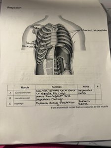
Muscles of Respiration
Overview
Muscles involved in respiration facilitate the movement of air into and out of the lungs.
External intercostals: Elevate ribs during inspiration (intercostal nerves).
Internal intercostals: Depress ribs during forced expiration (intercostal nerves).
Diaphragm: Flattens during inspiration, increases thoracic volume (phrenic nerve).
Muscle | Function | Nerve |
|---|---|---|
External intercostals | Elevate ribs during inspiration | Intercostal nerves |
Internal intercostals | Depress ribs during expiration | Intercostal nerves |
Diaphragm | Flattens during inspiration | Phrenic nerve |
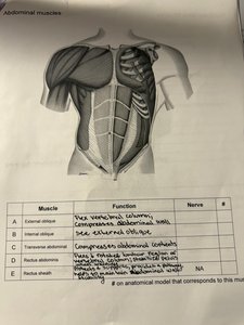
Abdominal Muscles
Overview
Abdominal muscles support the trunk, allow movement, and hold organs in place.
External oblique: Flexes vertebral column, compresses abdominal wall.
Internal oblique: Same as external oblique.
Transverse abdominis: Compresses abdominal contents.
Rectus abdominis: Flexes vertebral column.
Muscle | Function | Nerve |
|---|---|---|
External oblique | Flexes vertebral column, compresses abdominal wall | NA |
Internal oblique | Same as external oblique | NA |
Transverse abdominis | Compresses abdominal contents | NA |
Rectus abdominis | Flexes vertebral column | NA |
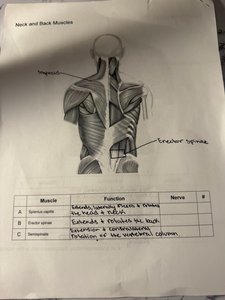
Neck and Back Muscles
Overview
These muscles are essential for posture and movement of the vertebral column.
Splenius capitis: Extends, rotates, and flexes the head and neck.
Erector spinae: Extends and rotates the back.
Semispinalis: Extension of the vertebral column.
Muscle | Function | Nerve |
|---|---|---|
Splenius capitis | Extends, rotates, flexes head/neck | NA |
Erector spinae | Extends, rotates back | NA |
Semispinalis | Extension of vertebral column | NA |
Summary Table: Axial Muscles and Their Functions
Region | Muscle | Function | Nerve |
|---|---|---|---|
Face | Orbicularis oculi | Closes eyelids | CN VII |
Jaw | Masseter | Elevates mandible | CN V |
Tongue | Genioglossus | Protrudes tongue | CN XII |
Neck | Sternocleidomastoid | Flexes head | CN XI |
Thorax | Diaphragm | Inspiration | Phrenic |
Abdomen | Rectus abdominis | Flexes vertebral column | NA |
Back | Erector spinae | Extends back | NA |
Additional info: Nerve supply is critical for muscle function; damage to these nerves can result in loss of movement or muscle tone. Axial muscles are distinct from appendicular muscles, which move the limbs.
