 Indietro
BIO 203 Unit 3 Study Guide: Nervous System Structure and Function
Guida di studio - Note intelligenti
 Appunti personalizzati basati sui tuoi materiali, ampliati con definizioni chiave, esempi e contesto.
Neural/Nervous Tissue and Neural/Nervous Physiology
Neuronal Communication and Neurotransmitters
The nervous system relies on specialized cells called neurons to transmit information throughout the body. Neurons communicate via electrical impulses and chemical signals known as neurotransmitters.
Neurotransmitters: Chemical messengers released at synapses. Examples include acetylcholine and dopamine.
Depolarization: If a post-synaptic neuron is depolarized, it is more likely to generate an action potential.
Hyperpolarization: Makes the neuron less likely to fire an action potential.
Example: Acetylcholine is released at neuromuscular junctions to stimulate muscle contraction.
Organization of the Nervous System
The nervous system is organized into central and peripheral divisions, each with distinct functions and structures.
Central Nervous System (CNS): Includes the brain and spinal cord.
Peripheral Nervous System (PNS): Comprises nerves and ganglia outside the CNS.
Flow Chart: Typically starts with CNS, branches to PNS, then divides into sensory (afferent) and motor (efferent) divisions.
Structure and Function of a Neuron
Neurons have specialized structures for receiving, processing, and transmitting information.
Cell body (soma): Contains the nucleus and organelles.
Dendrites: Receive information from other neurons.
Nissl bodies: Rough endoplasmic reticulum for protein synthesis.
Axon hillock: Initiates action potentials.
Axon: Conducts impulses away from the cell body.
Nodes of Ranvier: Gaps in myelin sheath for saltatory conduction.
Schwann cells: Produce myelin in the PNS.
Myelin: Insulates axons, speeds up impulse transmission.
Axon terminals: Release neurotransmitters.
Example: Dendrites receive signals, axon transmits them, and axon terminals release neurotransmitters.
Classification of Neurons
Neurons are classified structurally and functionally.
Structural: Multipolar, bipolar, unipolar.
Functional: Sensory (afferent), motor (efferent), interneurons.
Neuroglia/Glial Cells
Six types of glial cells support and protect neurons.
Astrocytes: Support and nourish neurons.
Oligodendrocytes: Myelinate CNS axons.
Microglia: Immune defense in CNS.
Ependymal cells: Produce cerebrospinal fluid (CSF).
Schwann cells: Myelinate PNS axons.
Satellite cells: Support PNS neuron cell bodies.
Membrane Channels and Potentials
Neurons use membrane channels to generate electrical signals.
Leak channels: Always open, do not use ATP.
Gated channels: Require energy; types include voltage-gated, ligand-gated, and mechanically-gated.
Resting Membrane Potential (RMP): Typically -70 mV; membrane is polarized.
Action Potential: Rapid change in membrane potential; uses voltage-gated channels; membrane depolarizes.
Local Potentials and Summation
Local potentials are graded changes in membrane potential that can trigger action potentials if summed.
Local potentials: Begin in dendrites or soma; use ligand-gated channels.
Summation: Multiple local potentials can combine to reach threshold and start an action potential.
Depolarization, Repolarization, Hyperpolarization
Changes in membrane potential are caused by ion movement.
Depolarization: Na+ influx.
Repolarization: K+ efflux.
Hyperpolarization: Excess K+ efflux or Cl- influx.
Example: On a graph, depolarization rises, repolarization falls, hyperpolarization dips below RMP.
Threshold and Refractory Periods
Action potentials require a threshold stimulus and are followed by refractory periods.
Threshold: Usually -55 mV; all-or-none phenomenon.
Absolute refractory period: No new action potential possible.
Relative refractory period: Action potential possible with stronger stimulus.
Chemical Synaptic Transmission
Neurons communicate at synapses using neurotransmitters.
Pre-synaptic neuron: Releases neurotransmitter.
Synaptic vesicles: Store neurotransmitter.
Synaptic cleft: Gap between neurons.
Ligand-gated channels: Respond to neurotransmitter.
Myelination and Conduction
Myelin increases conduction speed in neurons.
Continuous propagation: Occurs in unmyelinated axons.
Saltatory conduction: Occurs in myelinated axons; faster.
Excitatory and Inhibitory Post-Synaptic Potentials
EPSPs and IPSPs determine whether a neuron will fire an action potential.
EPSPs: Depolarize membrane; increase likelihood of action potential.
IPSPs: Hyperpolarize membrane; decrease likelihood of action potential.
The Central Nervous System: Brain and Cranial Nerves
Protection and Meninges
The brain is protected by physical and biochemical mechanisms.
Physical protection: Skull, meninges.
Biochemical protection: Blood-brain barrier.
Meninges: Pia mater (innermost), arachnoid mater, dura mater (outermost).
Brain Ventricles and Cerebrospinal Fluid (CSF)
The brain contains four ventricles filled with CSF, which cushions and nourishes neural tissue.
Ventricles: Lateral (2), third, fourth.
CSF production: Choroid plexus.
CSF flow: Ventricles → subarachnoid space → dural venous sinuses.
Functions of CSF: Protection, nutrient transport, waste removal.
Lobes and Cortices of the Brain
The brain is divided into lobes with specialized functions.
Frontal lobe: Motor functions.
Parietal lobe: Sensory functions.
Temporal lobe: Association and auditory functions.
Occipital lobe: Visual processing.
Primary motor cortex: Controls voluntary movement.
Primary sensory cortex: Processes sensory input.
Cerebral Cortex, Basal Nuclei, Ganglia, and Limbic System
The cerebral cortex is the outer layer of gray matter; basal nuclei and ganglia are involved in movement and coordination; the limbic system regulates emotions.
Cerebral cortex: Gray matter; higher processing.
Basal nuclei: Deep gray matter; movement regulation.
Ganglia: Clusters of neuron cell bodies in PNS.
Limbic system: Emotion and memory.
Diencephalon and Brainstem Functions
The diencephalon and brainstem control vital functions and relay information.
Thalamus: Sensory relay.
Hypothalamus: Homeostasis.
Midbrain, pons, medulla oblongata: Autonomic and reflex functions.
Reticular formation: Arousal and consciousness.
Memory Types
Memory is stored in various forms.
Immediate memory: Seconds.
Short-term memory: Minutes to hours.
Long-term memory: Days to years.
Cranial Nerves
There are twelve cranial nerves, each with specific functions.
Motor, sensory, mixed: Classification based on function.
Mnemonic: "Tale of Old Opie" for names; "Some say marry money..." for function.
The Central Nervous System: Spinal Cord and Peripheral Nervous System
Spinal Cord Structure and Function
The spinal cord transmits information between the brain and body and coordinates reflexes.
Decussation: Crossing of nerve fibers.
Cauda equina: Bundle of spinal nerves; clinical significance in nerve damage.
Somatic motor neuron cell bodies: Located in ventral horn.
Somatic sensory neuron cell bodies: Located in dorsal root ganglion.
Visceral motor impulses: Originate in lateral horn.
Spinal Nerve Tracts
Spinal nerve tracts are bundles of axons carrying information.
Ascending tracts: Sensory; e.g., spinocerebellar, spinothalamic.
Descending tracts: Motor; e.g., corticospinal, rubrospinal, reticulospinal.
Naming: Based on origin and destination.
Motor Neurons and Reflexes
Motor neurons are classified as upper or lower; reflexes are automatic responses.
Upper motor neurons: Begin in cortex, end in spinal cord.
Lower motor neurons: Begin in spinal cord, end in muscle.
Reflexes: Knee-jerk, cross extensor, withdrawal.
Connective Tissue Layers of Nerves
Nerves are protected by connective tissue layers.
Endoneurium: Covers individual axons.
Perineurium: Covers fascicles.
Epineurium: Covers entire nerve.
Spinal Nerves and Plexuses
Spinal nerves are mixed and organized into plexuses.
Mixed nerves: Carry both sensory and motor fibers.
31 pairs: Cervical, thoracic, lumbar, sacral.
Nerve plexuses: Networks of nerves; cervical, brachial, lumbar, sacral (lumbosacral).
Thoracic plexus: Not present; thoracic nerves form intercostal nerves.
Functions: Cervical (neck), brachial (arm), lumbar (leg), sacral (leg and foot).
Spinal Cord Cross-Section: Neuron Pathways
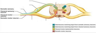
This diagram illustrates the pathways of somatic and visceral sensory and motor neurons in the spinal cord, showing their locations and connections.
Somatic sensory neurons: Enter dorsal horn.
Visceral sensory neurons: Enter dorsal horn.
Visceral motor neurons: Originate in lateral horn.
Somatic motor neurons: Originate in ventral horn.
Interneurons: Connect sensory and motor pathways.
Neuron Type | Location | Function |
|---|---|---|
Somatic sensory | Dorsal root ganglion | Receives sensory input from body |
Visceral sensory | Dorsal root ganglion | Receives sensory input from organs |
Visceral motor | Lateral horn | Controls autonomic functions |
Somatic motor | Ventral horn | Controls voluntary muscle movement |
Example: The knee-jerk reflex involves somatic sensory and motor neurons connecting through interneurons in the spinal cord.
Additional info: Academic context was added to clarify neuron types, spinal cord structure, and nerve plexus functions.
----------------------------------------
