 Indietro
Blood and Circulation: Structure, Function, and Physiology
Guida di studio - Note intelligenti
 Appunti personalizzati basati sui tuoi materiali, ampliati con definizioni chiave, esempi e contesto.
Overview of the Cardiovascular System
Introduction to the Cardiovascular System
The cardiovascular system is essential for transporting materials throughout the body, including nutrients, gases, hormones, and waste products. It consists of three main components: blood (the fluid), the heart (the pump), and blood vessels (the tubes).
Blood: Transports oxygen, carbon dioxide, nutrients, hormones, and waste products.
Heart: Pumps blood throughout the body, maintaining circulation.
Blood Vessels: Include arteries, veins, and capillaries, which serve as conduits for blood flow.
Functions:
Transport of materials from the external environment (nutrients, water, gases).
Distribution of materials between cells (hormones, immune cells, antibodies).
Removal of cellular waste (CO2, heat, metabolic waste).
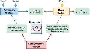
Components of Blood
Plasma
Plasma is the liquid extracellular matrix of blood, making up about 55% of total blood volume.
Water (92%): Solvent for carrying other substances.
Proteins (~7%): Includes albumins (transport), globulins (transport and immunity), fibrinogen (clotting), and transferrin (iron transport).
Other Solutes: Ions, organic molecules, gases, vitamins, and trace elements.
Formed Elements
The cellular components of blood are called formed elements and include:
Red Blood Cells (Erythrocytes): Carry oxygen and carbon dioxide.
White Blood Cells (Leukocytes): Immune defense.
Platelets (Thrombocytes): Involved in blood clotting.
Plasma Proteins Table
Protein | Main Function |
|---|---|
Albumins | Transport of substances |
Globulins | Transport and immunity |
Fibrinogen | Blood clotting |
Transferrin | Iron transport |
Hematopoiesis: Blood Cell Production
Definition and Regulation
Hematopoiesis is the process of blood cell production, occurring primarily in the red bone marrow. It is regulated by cytokines, including interleukins and specific growth factors.
Erythropoiesis: Production of red blood cells, stimulated by erythropoietin (EPO).
Thrombopoiesis: Production of platelets, stimulated by thrombopoietin (TPO).
Leukopoiesis: Production of white blood cells, stimulated by colony-stimulating factors (CSFs).
Red Blood Cells (Erythrocytes)
Structure and Function
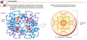
Mature erythrocytes lack a nucleus and mitochondria, optimizing them for gas transport. Their main protein, hemoglobin, binds oxygen and carbon dioxide.
Hemoglobin: Each molecule consists of four protein chains, each with a heme group that binds oxygen.
Lifespan: Approximately 120 days.
Recycling: Iron from heme is reused; heme is broken down into bilirubin for excretion.
Hematocrit: The ratio of red blood cells to plasma, expressed as a percentage.
RBC Disorders
Accelerated RBC Loss: Due to blood loss or hemolytic anemias (e.g., sickle cell anemia, malaria).
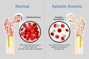
Decreased RBC Production: Caused by bone marrow failure (aplastic anemia), iron deficiency, folic acid or vitamin B12 deficiency, or inadequate erythropoietin production.
Platelets (Thrombocytes)
Structure and Function
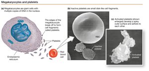
Platelets are cell fragments derived from megakaryocytes. They play a crucial role in hemostasis, immunity, and inflammation.
Origin: Produced in bone marrow from megakaryocytes.
Contents: Mitochondria, smooth endoplasmic reticulum, and granules containing cytokines and growth factors.
Lifespan: About 10 days.
Hemostasis and Coagulation
Stages of Hemostasis
Hemostasis is the process that prevents blood loss from damaged vessels and occurs in three stages:
Vasoconstriction: Chemicals released from the injury site cause vessel constriction.
Platelet Plug Formation: Platelets adhere to exposed collagen and aggregate to form a temporary plug.
Coagulation: Formation of a stable blood clot (thrombus) via the coagulation cascade.
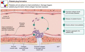
Coagulation Pathways
Coagulation involves two pathways (intrinsic and extrinsic) that converge on a common pathway, resulting in the conversion of fibrinogen to fibrin by thrombin.
Fibrinolysis: The breakdown of the fibrin clot by plasmin, which is activated from plasminogen by tissue plasminogen activator (tPA).
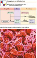
Anticoagulants and Hemophilia
Anticoagulants: Substances that prevent excessive clotting, including endogenous molecules like heparin and antithrombin.
Hemophilia: A group of disorders where one or more factors in the coagulation cascade are defective or missing, leading to impaired clotting.
Leukocytes (White Blood Cells)
Types and Functions
Leukocytes are immune cells capable of leaving the circulation (diapedesis) and migrating to sites of infection or injury.
Eosinophils: Reduce inflammation and combat parasitic infections.
Basophils: Mediate inflammation (release histamine) and prevent clotting (release heparin); called mast cells in tissues.
Neutrophils: First responders to bacterial invasion.
Lymphocytes: Produce antibodies and identify pathogens.
Monocytes: Phagocytic cells that present antigens to lymphocytes.

The Heart
Structure and Circulation
The heart is divided by a septum into left and right halves. Each half contains an atrium (receives blood) and a ventricle (pumps blood out).
Pulmonary Circulation: Right side pumps blood to the lungs for oxygenation.
Systemic Circulation: Left side pumps oxygenated blood to the body.
Heart Valves
Heart valves ensure one-way flow of blood:
Atrioventricular (AV) Valves: Tricuspid (right), Bicuspid/Mitral (left).
Semilunar Valves: Aortic and pulmonary valves, between ventricles and arteries.
Core Concept: Gradients and Flow
Pressure Gradients
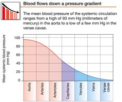
Blood flows from regions of high pressure to low pressure. The pressure gradient () drives blood through the circulatory system.
Highest pressure: Aorta
Lowest pressure: Venae cavae
Cardiac Conduction System
Autorhythmic Cells and Pacemaker Activity
The heart has its own conduction system, independent of nervous system input.
Sinoatrial (SA) Node: Pacemaker cells spontaneously depolarize to initiate the heartbeat.
Atrioventricular (AV) Node: Slows conduction, allowing atrial contraction before ventricular contraction.
AV Bundle, Bundle Branches, Purkinje Fibers: Rapidly conduct electrical signals to ventricles.
Action Potentials in Cardiac Autorhythmic Cells
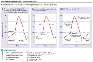
Pacemaker potential: Spontaneous depolarization due to opening of Na+/K+ (If or HCN) channels.
At threshold (-40 mV), Ca2+ channels open, leading to rapid depolarization.
Action Potentials in Contractile Cardiac Muscle Cells
Phases of the Action Potential
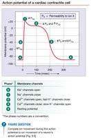
Cardiac muscle action potentials differ from skeletal muscle due to a prolonged plateau phase, preventing tetanus.
Depolarization: Na+ channels open.
Initial Repolarization: Na+ channels close, some K+ channels open.
Plateau: Ca2+ channels open, some K+ channels close.
Rapid Repolarization: Ca2+ channels close, K+ channels open.
Resting Membrane Potential: Restored by K+ permeability.
Calcium-Induced Calcium Release
The action potential triggers Ca2+ entry, which induces further Ca2+ release from the sarcoplasmic reticulum, initiating contraction.
Cardiac Muscle Relaxation
Relaxation occurs as Ca2+ is pumped back into the sarcoplasmic reticulum and out of the cell via the Na+-Ca2+ exchanger.
Refractory Periods and Summation
Prevention of Tetanic Contractions
Cardiac muscle has a long refractory period, preventing summation and tetanus, which is essential for rhythmic heart function. 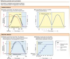
The Cardiac Cycle
Phases of the Cardiac Cycle
The cardiac cycle consists of alternating periods of contraction (systole) and relaxation (diastole).
Atrial and Ventricular Diastole: Heart at rest; blood flows into atria and ventricles.
Atrial Systole: Atria contract, completing ventricular filling (end-diastolic volume, EDV).
Early Ventricular Contraction: AV valves close (first heart sound, "Lub"), isovolumic contraction.
Ventricular Ejection: Semilunar valves open, blood is ejected into arteries.
Ventricular Relaxation: Semilunar valves close (second heart sound, "Dup"), isovolumic relaxation.
Key Terms:
Isovolumic Contraction/Relaxation: Periods when all valves are closed and ventricular volume does not change.
End-Diastolic Volume (EDV): Volume of blood in ventricle at the end of relaxation.
Summary Table: Blood Components and Functions
Component | Main Function |
|---|---|
Plasma | Transport of nutrients, hormones, waste |
Red Blood Cells | Oxygen and carbon dioxide transport |
White Blood Cells | Immune defense |
Platelets | Blood clotting |
Review Questions
What are the components of blood and their functions?
How does hemostasis occur?
How does the heart function?
How are cardiac muscle action potentials different from skeletal muscle action potentials?
Describe the phases of the cardiac cycle.
