 Indietro
Blood Flow and Pressure: Structure, Function, and Regulation
Guida di studio - Note intelligenti
 Appunti personalizzati basati sui tuoi materiali, ampliati con definizioni chiave, esempi e contesto.
Blood Vessels: Structure and Function
Types of Blood Vessels
The cardiovascular system consists of a network of vessels that transport blood throughout the body. These vessels are classified based on their structure and function:
Arteries: Carry blood away from the heart. Subtypes include elastic arteries (conducting vessels), muscular arteries (distributing vessels), and arterioles (resistance vessels).
Capillaries: Smallest vessels, primary site of exchange between blood and interstitial fluid.
Veins: Carry blood toward the heart, act as capacitance vessels, and contain valves to prevent backflow.
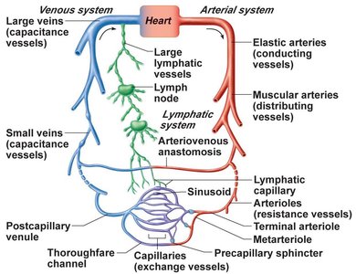
Structural Differences Among Arteries, Veins, and Capillaries
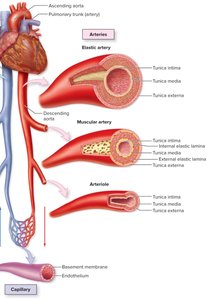
Blood vessels differ in their wall composition and function:
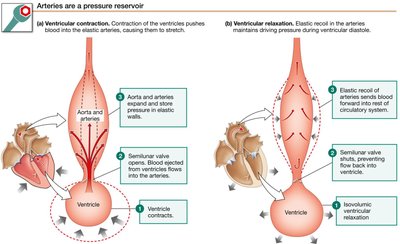
Elastic arteries: Thick tunica media with abundant elastic fibers, near the heart, act as pressure reservoirs.
Muscular arteries: More smooth muscle, less elastic tissue, regulate blood distribution.
Arterioles: Small diameter, major site of resistance, regulate blood flow to capillaries.
Veins: Thin walls, large lumen, contain valves, act as blood reservoirs.
Capillaries: Single layer of endothelium, lack smooth muscle, facilitate exchange.
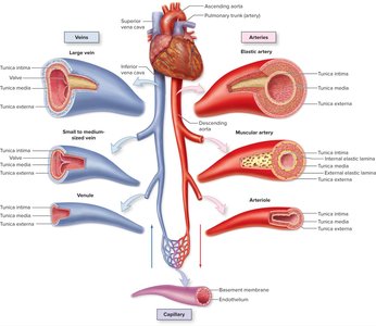
Branching and Distribution
Blood vessels branch extensively, increasing total cross-sectional area and decreasing pressure as blood moves away from the heart. The aorta branches into thousands of arteries, millions of arterioles, and billions of capillaries.
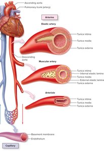
Capillary Beds and Blood Flow Regulation
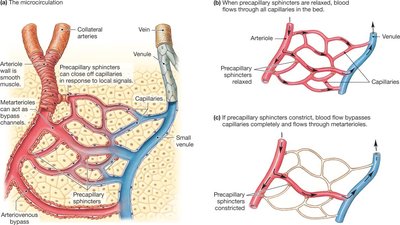
Capillary Beds and Precapillary Sphincters
Capillary beds are networks of capillaries supplied by arterioles. Precapillary sphincters regulate blood flow into capillaries, influenced by the sympathetic nervous system and local metabolites.
Bulk flow: Movement of ions and water.
Simple diffusion: Movement of gases and lipids.
Facilitated diffusion: Movement of water via aquaporins.
Types of Capillaries
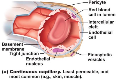
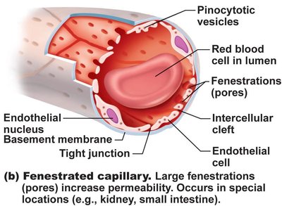
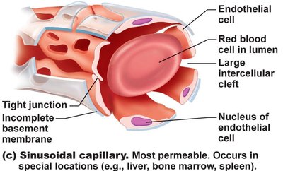
Capillaries are classified based on permeability:
Continuous capillaries: Most common, tight junctions, least permeable, found in skin and muscle.
Fenestrated capillaries: Have pores, increased permeability, found in kidneys, small intestine, endocrine glands.
Sinusoidal capillaries: Most permeable, large gaps, found in liver, spleen, bone marrow.
Veins: Blood Reservoirs and Return to the Heart
Vein Structure and Function
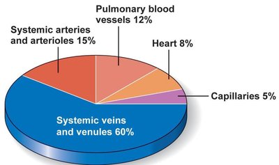
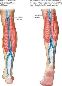
Veins act as blood reservoirs, holding up to 65% of blood at rest. They have thin walls, low pressure, and valves to prevent backflow. The muscular and thoracic pumps aid venous return.
Blood Pressure and Flow
Pressure Gradients and Flow
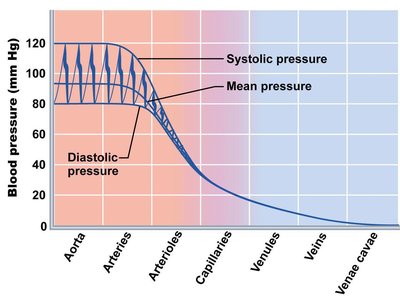
Blood flows from regions of high to low pressure, driven by the pressure gradient (ΔP). Fluid motion includes dynamic (kinetic energy) and lateral (hydrostatic pressure) components. Pressure decreases with distance due to friction.
Systolic pressure: Peak pressure during heart contraction (120 mmHg).
Diastolic pressure: Minimum pressure during heart relaxation (80 mmHg).
Pulse pressure: Difference between systolic and diastolic.
Mean arterial pressure (MAP): Average pressure driving blood flow.
MAP Calculation:
MAP = diastolic + (pulse pressure/3)
MAP = (2/3 × diastolic) + (1/3 × systolic)
Resistance and Flow: Poiseuille’s Law
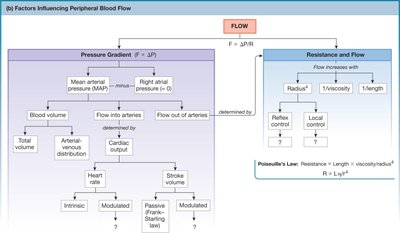
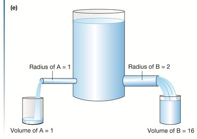
Resistance (R) opposes blood flow. The radius of a vessel has the greatest impact on resistance. Poiseuille’s Law describes the relationship:
Blood Flow (F):
Resistance:
Cardiac Output, Peripheral Resistance, and MAP
Determinants of Mean Arterial Pressure (MAP)
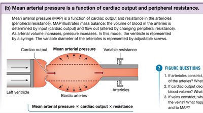
MAP is determined by cardiac output (CO) and peripheral resistance (R):
MAP:
Cardiac Output:
Stroke Volume:
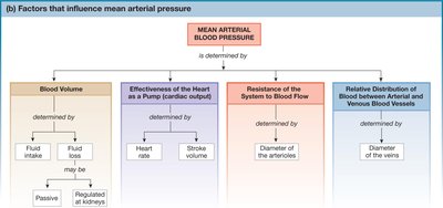
Blood Pressure Regulation
Short-Term Regulation: Baroreceptor Reflex
Baroreceptors in arteries detect changes in blood pressure and signal the medulla to adjust sympathetic activity:
High BP: Decreased sympathetic activity → vasodilation, lower heart rate, lower CO.
Low BP: Increased sympathetic activity → vasoconstriction, higher heart rate, higher CO.
MAP Equation:
Long-Term Regulation: Kidney Function
The kidneys regulate blood volume and pressure by controlling water and sodium balance. Fluid intake or intravenous infusion can correct low blood volume.
Distribution of Blood to Tissues
Blood Distribution and Tissue Perfusion
Blood distribution varies according to the metabolic needs of tissues, governed by local and autonomic mechanisms. Arterioles are arranged in parallel, allowing individual regulation of flow.
Intrinsic mechanisms: Local metabolites (O2, CO2, H+, K+, prostaglandins, adenosine, nitric oxide) trigger vasodilation.
Extrinsic mechanisms: Neuronal and hormonal control.
Capillary Exchange
Mechanisms of Exchange
Capillary exchange occurs via:
Bulk flow: Balances fluid volume between interstitial fluid and blood.
Hydrostatic pressure: Due to blood pressure in vessels relative to interstitial fluid.
Osmotic pressure: Due to plasma proteins (mainly albumin).
Summary Table: Comparison of Blood Vessel Types
Type | Structure | Function | Permeability |
|---|---|---|---|
Elastic Artery | Thick tunica media, elastic fibers | Pressure reservoir | Low |
Muscular Artery | More smooth muscle, less elastic | Distributes blood | Low |
Arteriole | Small diameter, muscular | Regulates flow/resistance | Low |
Capillary (Continuous) | Endothelium, tight junctions | Exchange | Least |
Capillary (Fenestrated) | Endothelium, pores | Exchange | Moderate |
Capillary (Sinusoidal) | Large gaps, incomplete membrane | Exchange | Most |
Vein | Thin wall, valves | Reservoir, return | Low |
Additional info: Academic context was added to clarify vessel types, regulatory mechanisms, and formulas for blood flow and pressure.
