 Indietro
Blood: Leukocytes, Platelets, and Hemostasis
Guida di studio - Note intelligenti
 Appunti personalizzati basati sui tuoi materiali, ampliati con definizioni chiave, esempi e contesto.
Leukocytes: Structure and Function
General Characteristics of Leukocytes
Leukocytes, also known as white blood cells (WBCs), are the only formed elements in blood that are complete cells, possessing nuclei and organelles. They constitute less than 1% of total blood volume and play a crucial role in defending the body against disease. Leukocytes can exit capillaries through a process called diapedesis and move through tissue spaces by amoeboid motion and positive chemotaxis.
Granulocytes: Contain visible cytoplasmic granules.
Agranulocytes: Lack visible cytoplasmic granules.
Mnemonic for decreasing abundance: Never let monkeys eat bananas.
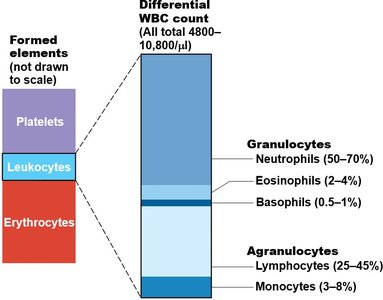
Types and Relative Percentages of Leukocytes
Leukocytes are classified into granulocytes and agranulocytes, each with distinct types and relative percentages in normal blood:
Granulocytes: Neutrophils (50–70%), Eosinophils (2–4%), Basophils (0.5–1%)
Agranulocytes: Lymphocytes (25–45%), Monocytes (3–8%)
Granulocytes
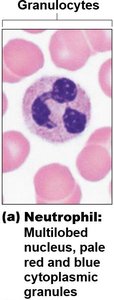
Neutrophils
Neutrophils are the most numerous WBCs, accounting for 50–70% of all leukocytes. They are about twice the size of red blood cells and are highly phagocytic, often referred to as "bacterial slayers." Neutrophils kill microbes through a process called respiratory burst, synthesizing potent oxidizing substances such as bleach or hydrogen peroxide.
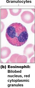
Eosinophils
Eosinophils make up 2–4% of leukocytes. They play a role in allergies, asthma, immune response modulation, and defense against parasitic infections.
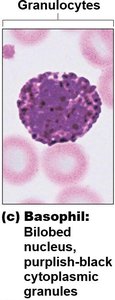
Basophils
Basophils are the rarest WBCs, accounting for only 0.5–1% of leukocytes. Their large, purplish-black granules contain histamine, an inflammatory chemical that acts as a vasodilator and attracts other WBCs to inflamed sites.
Agranulocytes
Lymphocytes
Lymphocytes are the second most numerous WBC, accounting for about 25% of leukocytes. They are mostly found in lymphoid tissues such as lymph nodes and spleen and are crucial to immunity. There are two main types:
T lymphocytes (T cells): Act against virus-infected cells and tumor cells.
B lymphocytes (B cells): Give rise to plasma cells, which produce antibodies.
Monocytes
Monocytes are the largest leukocytes, accounting for 3–8% of WBCs. They leave circulation, enter tissues, and differentiate into macrophages, which are actively phagocytic and crucial against viruses, intracellular bacterial parasites, and chronic infections. Monocytes also activate lymphocytes to mount an immune response.
Production and Life Span of Leukocytes
Leukopoiesis
Leukopoiesis is the production of WBCs, stimulated by chemical messengers from red bone marrow and mature WBCs:
Interleukins: Numbered (e.g., IL-3, IL-5).
Colony-stimulating factors (CSFs): Named for the WBC type they stimulate (e.g., granulocyte-CSF).
All leukocytes originate from hemocytoblast stem cells that branch into two pathways:
Lymphoid stem cells: Produce lymphocytes.
Myeloid stem cells: Produce all other elements.
Platelets
Structure and Function
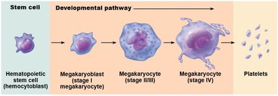
Platelets are cytoplasmic fragments of megakaryocytes, with blue-staining outer regions and purple granules. These granules contain serotonin, enzymes, ADP, and platelet-derived growth factor (PDGF). Platelets play a key role in the clotting process, forming a temporary platelet plug to seal breaks in blood vessels. Normal platelet count is 150,000–400,000 platelets/ml of blood.
Platelet Formation
Platelet formation is regulated by thrombopoietin. Platelets are formed in the myeloid line from megakaryoblasts. Megakaryocytes send cytoplasmic projections into the lumen of capillaries, which break off into platelet fragments. Platelets age quickly and degenerate in about 10 days.
Hemostasis
Overview of Hemostasis
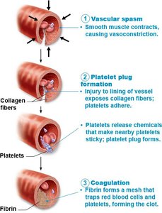
Hemostasis is a rapid series of reactions that stop bleeding, requiring clotting factors and substances released by platelets and injured tissues. The process involves three steps:
Vascular spasm: Vasoconstriction in response to injury.
Platelet plug formation: Platelets adhere to exposed collagen fibers and release chemicals to attract more platelets.
Coagulation: Blood clotting reinforces the platelet plug with fibrin threads, transforming blood from liquid to gel.
Step 1: Vascular Spasm
Triggered by direct injury to vascular smooth muscle, chemicals released by endothelial cells and platelets, and pain reflexes.
Most effective in smaller blood vessels.
Reduces blood flow until other mechanisms activate.
Step 2: Platelet Plug Formation
Platelets stick to collagen fibers exposed by vessel damage.
Intact vessel walls prevent platelet sticking via prostacyclins and nitric oxide.
Activated platelets swell, become spiked and sticky, and release ADP, serotonin, and thromboxane A2.
Positive feedback cycle: more platelets stick and release chemicals, attracting more platelets.
Step 3: Coagulation
Reinforces platelet plug with fibrin threads.
Blood is transformed from liquid to gel, effectively sealing larger vessel breaks.
Blood Transfusions
Minimizing Effects of Blood Loss
Cardiovascular system reduces volume of affected blood vessels and increases RBC production.
Body can compensate for only limited blood loss (15–30% causes pallor and weakness).
Type O: Universal donor (no A or B antigens).
Type AB: Universal recipient (no anti-A or anti-B antibodies).
Summary Table: Leukocyte Types and Functions
Type | Relative Percentage | Main Function |
|---|---|---|
Neutrophils | 50–70% | Phagocytosis; bacterial defense |
Eosinophils | 2–4% | Allergy, asthma, parasite defense |
Basophils | 0.5–1% | Release histamine; inflammation |
Lymphocytes | 25–45% | Immunity (T cells, B cells) |
Monocytes | 3–8% | Phagocytosis; chronic infection defense |
Key Equations
Platelet Count Formula
Normal platelet count:
White Blood Cell Differential Formula
WBC differential count:
Hemostasis Steps (Summary)
Hemostasis:
----------------------------------------
