 Indietro
Blood: Structure, Function, and Clinical Significance
Guida di studio - Note intelligenti
 Appunti personalizzati basati sui tuoi materiali, ampliati con definizioni chiave, esempi e contesto.
Blood: Structure, Function, and Clinical Significance
Functions of Blood
Blood is a vital connective tissue that serves as the internal transport system of the body. Its functions are categorized into transport, regulation, and protection.
Transport: Delivers oxygen (O2) and nutrients to cells, removes metabolic wastes (to lungs and kidneys), and transports hormones from endocrine organs to target tissues.
Regulation: Maintains body temperature, normal pH (using buffers and bicarbonate ions), and adequate fluid volume in the circulatory system.
Protection: Prevents blood loss (via clot formation) and infection (through antibodies, complement proteins, and white blood cells).
Components of Blood
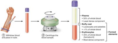
Blood consists of plasma (liquid matrix) and formed elements (cells and cell fragments). It is classified as connective tissue due to its cellular and extracellular components.
Plasma: Straw-colored fluid (~90% water) containing nutrients, gases, hormones, wastes, proteins, and inorganic ions. Plasma proteins (mainly albumin) regulate osmotic pressure and pH.
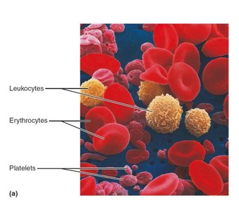
Formed Elements: Include erythrocytes (RBCs), leukocytes (WBCs), and platelets. RBCs lack nuclei and most organelles; platelets are cell fragments; WBCs are complete cells.
Physical Characteristics and Volume
Blood is a viscous, opaque fluid with a metallic taste. Its color varies with oxygen content: bright red when oxygen-rich, dark red when oxygen-poor. Blood pH is tightly regulated between 7.35–7.45, and it constitutes about 8% of body weight (5–6 L in males, 4–5 L in females).
Formed Elements: Erythrocytes, Leukocytes, Platelets
The formed elements have unique features and functions:
Erythrocytes (RBCs): Specialized for gas transport; lack nuclei and most organelles.
Leukocytes (WBCs): Complete cells involved in immune defense.
Platelets: Cell fragments essential for clotting.
Erythrocytes: Structure and Function
Structural Characteristics
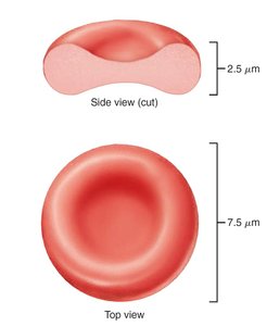
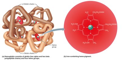
Erythrocytes are small, biconcave cells (~7.5 μm diameter) lacking nuclei and most organelles. Their shape provides a large surface area-to-volume ratio, facilitating efficient gas exchange. They are filled with hemoglobin (Hb), the protein responsible for oxygen and carbon dioxide transport.
Biconcave shape: Maximizes surface area for diffusion.
High hemoglobin content: 97% of cell volume (excluding water).
Anaerobic metabolism: No mitochondria; ATP produced without consuming transported oxygen.
Hemoglobin Structure and Function
Hemoglobin is a complex protein composed of four globin chains (two alpha, two beta) and four heme groups, each with a central iron ion. Each iron binds one oxygen molecule, allowing each hemoglobin to carry four oxygen molecules.
Oxygen transport: Hb binds reversibly with oxygen; normal values: males 13–18g/100ml, females 12–16g/100ml.
Color: Oxygen-bound Hb (oxyhemoglobin) is bright red; deoxygenated Hb (deoxyhemoglobin) is dark red.
Carbon dioxide transport: 20% of CO2 binds to Hb, forming carbaminohemoglobin.
Erythropoiesis: Production and Regulation
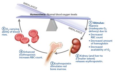
Hematopoiesis (formation of blood cells) occurs in red bone marrow. Erythropoiesis is regulated by erythropoietin (EPO), a hormone released by the kidneys in response to hypoxia (low oxygen levels).
Dietary requirements: Amino acids, lipids, carbohydrates, vitamin B12, folic acid, and iron are essential for RBC production.
Iron: Most found in hemoglobin; stored as ferritin and hemosiderin; transported by transferrin.
Life Cycle and Destruction of Erythrocytes
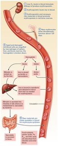
RBCs have a lifespan of 100–120 days. Old RBCs are destroyed by macrophages in the spleen; their components are recycled or excreted.
Iron: Stored for reuse.
Heme: Degraded to bilirubin, excreted in bile, converted to urobilinogen and stercobilin (feces pigment).
Globin: Broken down to amino acids.
Erythrocyte Disorders
Disorders are classified as anemia (insufficient RBCs or Hb) or polycythemia (excess RBCs).
Anemia: Causes include blood loss, insufficient RBC production, or excessive RBC destruction. Types include iron-deficiency, pernicious, renal, aplastic, hemolytic, thalassemia, and sickle-cell anemia.
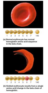
Sickle-cell anemia: Genetic mutation causes abnormal HbS, leading to sickled RBCs that rupture easily and block vessels.
Polycythemia: Excess RBCs increase blood viscosity; causes include bone marrow cancer (polycythemia vera), high altitude, or blood doping.
Leukocytes: Structure and Function
General Characteristics
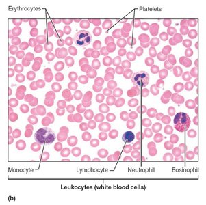
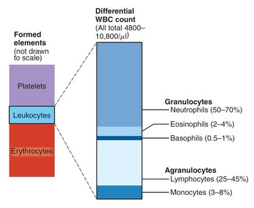
Leukocytes (WBCs) are complete cells with nuclei and organelles. They defend the body against pathogens and are classified as granulocytes or agranulocytes.
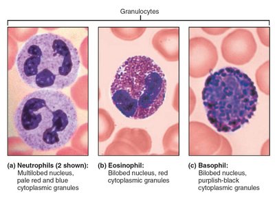
Granulocytes: Neutrophils, eosinophils, basophils (contain cytoplasmic granules).
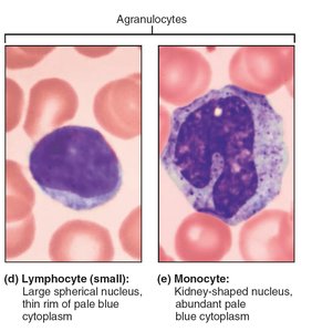
Agranulocytes: Lymphocytes, monocytes (lack visible granules).
Leukocytosis: Elevated WBC count (>11,000/μL) is a normal response to infection.
Granulocytes
Neutrophils: Most numerous; phagocytic; kill bacteria with defensins and oxidizing substances.
Eosinophils: Attack parasitic worms; modulate immune responses; involved in allergies and asthma.
Basophils: Rarest; release histamine (inflammatory mediator); similar to mast cells.
Agranulocytes
Lymphocytes: Second most numerous; crucial for immunity. T cells attack infected/tumor cells; B cells produce antibodies.
Monocytes: Largest WBCs; differentiate into macrophages; active in phagocytosis and chronic infections.
Leukopoiesis: Production and Life Span
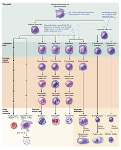
Leukopoiesis is stimulated by interleukins and colony-stimulating factors (CSFs). Hematopoietic stem cells differentiate into myeloid or lymphoid lineages, producing various WBCs.
Granulocyte production: Myeloblast → promyelocyte → myelocyte → band cell → mature granulocyte.
Lymphocyte production: Lymphoid stem cell → lymphocyte.
Leukocyte Disorders
Leukopenia: Low WBC count; often drug-induced.
Leukemias: Cancerous overproduction of abnormal WBCs; classified as acute/chronic and myeloid/lymphocytic.
Infectious mononucleosis: Viral disease (Epstein-Barr); excess atypical lymphocytes.
Platelets: Structure and Function
Platelet Formation and Role
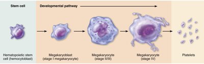
Platelets are cell fragments from megakaryocytes, essential for blood clotting. Platelet formation (thrombopoiesis) is regulated by thrombopoietin.
Formation: Megakaryocytes fragment in bone marrow and lungs, releasing platelets into circulation.
Function: Form temporary plugs to seal vessel breaks; contain chemicals for clotting.
Hemostasis: Prevention of Blood Loss
Steps of Hemostasis
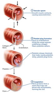
Hemostasis is a rapid, localized process to stop bleeding, involving three steps:
Vascular spasm: Vasoconstriction reduces blood loss.
Platelet plug formation: Platelets adhere to exposed collagen, become activated, and aggregate.
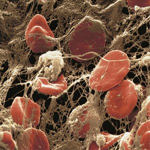
Coagulation: Clotting factors form a fibrin mesh, stabilizing the plug.
Coagulation Pathways
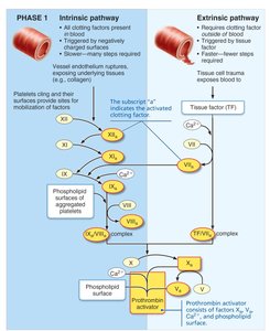
Coagulation occurs via intrinsic and extrinsic pathways, both leading to the formation of prothrombin activator and ultimately a fibrin clot.
Intrinsic pathway: Triggered by factors within blood.
Extrinsic pathway: Triggered by tissue factor outside blood; faster.
Vitamin K: Required for synthesis of several clotting factors.
Clot Retraction and Fibrinolysis
After vessel repair, the clot is stabilized and removed:
Clot retraction: Platelet actin and myosin contract, pulling vessel edges together.
Fibrinolysis: Plasminogen is activated to plasmin, which digests fibrin and dissolves the clot.
Disorders of Hemostasis
Thromboembolic disorders: Undesirable clot formation (thrombus, embolus).
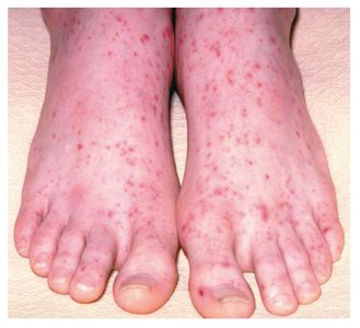
Bleeding disorders: Platelet or clotting factor deficiencies (thrombocytopenia, hemophilia, impaired liver function).
Disseminated intravascular coagulation (DIC): Both widespread clotting and severe bleeding.
Blood Transfusion and Blood Typing
Blood Groups and Compatibility
Transfusions require careful matching of blood groups to prevent fatal reactions. The ABO and Rh systems are most clinically significant.
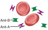
ABO system: Based on presence/absence of A and B antigens; plasma contains anti-A and/or anti-B antibodies.
Rh system: Rh+ (D antigen present); Rh- individuals can develop anti-Rh antibodies after exposure.
Universal donor: Type O; Universal recipient: Type AB.
Blood Group | RBC Antigens | Plasma Antibodies | Blood Received |
|---|---|---|---|
AB | A, B | None | A, B, AB, O |
B | B | Anti-A | B, O |
A | A | Anti-B | A, O |
O | None | Anti-A, Anti-B | O |
Blood Typing
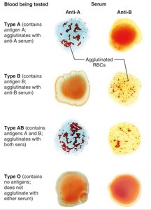
Blood is mixed with antibodies against A and B antigens; agglutination indicates presence of the antigen. Cross-matching ensures compatibility between donor and recipient.
Blood Tests and Developmental Aspects
Clinical Blood Tests
Hematocrit: Measures RBC proportion; low values indicate anemia.
Blood glucose: Screens for diabetes.
Differential WBC count: Assesses proportions of WBC types for diagnosis.
Prothrombin time (PT): Assesses clotting ability.
Complete blood count (CBC): Evaluates formed elements, hematocrit, hemoglobin.
Developmental Aspects
Fetal blood: Forms in yolk sac, liver, spleen; red bone marrow becomes primary site by seventh month.
Hemoglobin F: Higher affinity for oxygen than adult hemoglobin A.
Blood diseases of aging: Chronic leukemias, anemias, clotting disorders often linked to cardiovascular or immune system disorders.
