 Indietro
Blood Vessels and Circulatory Pathways: Control, Exchange, and Clinical Relevance
Guida di studio - Note intelligenti
 Appunti personalizzati basati sui tuoi materiali, ampliati con definizioni chiave, esempi e contesto.
The Cardiovascular System: Blood Vessels
Control of Blood Flow
The regulation of blood flow through body tissues, known as tissue perfusion, is essential for delivering oxygen and nutrients, removing wastes, facilitating gas exchange, absorbing nutrients, and forming urine. The rate of blood flow is tightly controlled to match the functional needs of each tissue or organ.
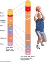
Key Point 1: Blood flow is redistributed during exercise, with skeletal muscles receiving a much larger proportion of total blood flow (from ~20% at rest to over 70% during strenuous activity).
Key Point 2: Intrinsic controls (local dilation of arterioles) increase blood flow to active muscles, while extrinsic controls decrease flow to less active organs.
Key Point 3: Mean arterial pressure (MAP) is maintained even as blood flow is redirected.
Example: During exercise, blood is shunted away from kidneys and digestive organs to supply working muscles.
Autoregulation: Intrinsic Regulation of Blood Flow
Autoregulation refers to local mechanisms that adjust blood flow to meet tissue needs. Two main types of intrinsic mechanisms determine the final autoregulatory response:
Metabolic controls: Increased metabolic activity leads to decreased O2 and increased metabolic byproducts (H+, K+), causing vasodilation via direct relaxation of arterioles and release of nitric oxide (NO).
Myogenic controls: Vascular smooth muscle responds to changes in MAP. Increased stretch (high MAP) causes constriction, while reduced stretch (low MAP) causes dilation, maintaining constant perfusion.
Long-term autoregulation: When short-term mechanisms are insufficient, angiogenesis (growth of new vessels) and enlargement of existing vessels occur over weeks or months.
Blood Flow in Special Areas
Skeletal Muscles
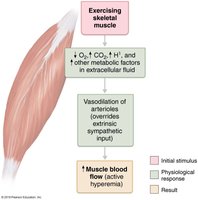
Blood flow to skeletal muscles varies with fiber type and activity. At rest, flow is maintained by myogenic and neural mechanisms (~1 L/min). During activity, active hyperemia increases flow up to tenfold, directly proportional to metabolic activity.
Example: Exercise increases O2 demand, CO2, and H+, triggering vasodilation and increased muscle blood flow.
Brain
Constant blood flow (~750 ml/min) is critical for brain function. Metabolic controls (decreased pH or increased CO2) cause vasodilation. Myogenic controls adjust vessel diameter in response to MAP changes. Extreme MAP values (<60 mm Hg or >160 mm Hg) can cause fainting or cerebral edema.
Skin
Blood flow through the skin supplies nutrients, regulates temperature, and serves as a reservoir. Flow is controlled by sympathetic reflexes and varies with temperature. Dilation occurs with heat, constriction with cold, affecting heat loss and retention.
Lungs
The pulmonary circuit is short, with arteries and arterioles resembling veins. Pulmonary arterial pressure is much lower (~24/10 mm Hg) than systemic (~120/80 mm Hg).
Heart
Coronary blood flow is influenced by aortic pressure and ventricular activity. During systole, vessels are compressed and flow ceases; during diastole, high aortic pressure restores flow. Cardiac cells extract 65% of delivered O2, so increased flow is necessary during exercise.
Capillary Exchange
Velocity of Blood Flow
Blood velocity changes throughout systemic circulation: fastest in the aorta, slowest in capillaries, and increases in veins. The speed is inversely related to total cross-sectional area, allowing slow capillary flow for efficient exchange.
Capillary Exchange of Respiratory Gases and Nutrients
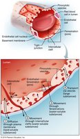
Molecules move between blood and interstitial fluid by diffusion, following concentration gradients. Four main routes exist for crossing capillary walls:
Direct diffusion through endothelial membranes (lipid-soluble molecules, e.g., O2, CO2)
Passage through intercellular clefts (water-soluble solutes)
Passage through fenestrations (pores in capillary walls)
Active transport via pinocytotic vesicles (large molecules, e.g., proteins)
Fluid Movements: Bulk Flow
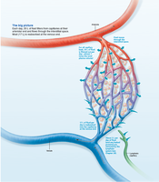
Bulk flow across capillary walls mixes plasma and interstitial fluid, maintaining the interstitial environment. Fluid is forced out at the arterial end and returns at the venous end. The direction and amount of flow depend on hydrostatic and osmotic pressures.
Capillary hydrostatic pressure (HPc): Forces fluid out at the arterial end (filtration), and in at the venous end (reabsorption). HPc is higher at the arterial end (35 mm Hg) than at the venous end (17 mm Hg).
Interstitial fluid hydrostatic pressure (HPif): Usually assumed to be zero due to lymphatic drainage.
Clinical Relevance: Edema
Homeostatic Imbalance: Edema
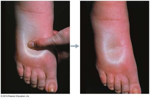
Edema is an abnormal increase in interstitial fluid, caused by increased outward pressure or decreased inward pressure. It may result from incompetent venous valves, vessel blockage, heart failure, or high blood volume. Inflammation increases capillary permeability, allowing proteins to leak and drawing fluid into the interstitial space.
Decreased lymphatic drainage (due to disease or surgery) can also cause edema.
Pitting edema is characterized by a visible indentation after pressure is applied to the skin.
Edema impairs tissue function by increasing diffusion distance for gases, nutrients, and wastes.
Circulatory Pathways: Blood Vessels of the Body
Pulmonary and Systemic Circulation
The vascular system consists of two main circulations:
Pulmonary circulation: Short loop from heart to lungs and back.
Systemic circulation: Long loop to all body parts and back.
The heart pumps blood via the aorta; blood returns via the superior and inferior vena cava and coronary sinus.
Differences Between Systemic Arteries and Veins
Arteries run deep; veins are both deep and superficial.
Deep veins share names with corresponding arteries; superficial veins do not.
Arteries and veins often run side by side with nerves.
Systemic vessels may not match on right and left sides; venous pathways are more interconnected and may have multiple names.
Major Arteries of the Systemic Circulation
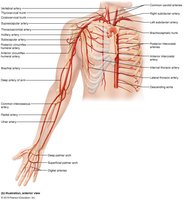
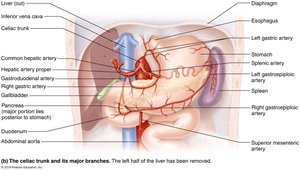
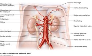
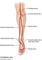
Systemic arteries deliver oxygenated blood throughout the body. The aorta branches into major arteries supplying the head, neck, upper limbs, thorax, abdomen, pelvis, and lower limbs.
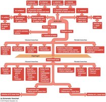
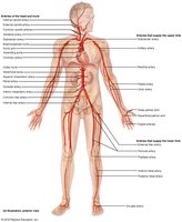
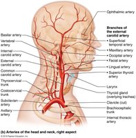
Arteries of the Head, Neck, and Brain
Arteries of the Right Upper Limb and Thorax
Arteries of the Abdomen
Arteries of the Right Pelvis and Lower Limb
Major Veins of the Systemic Circulation
Systemic veins return deoxygenated blood to the heart. The venous system is highly interconnected, with major veins draining the head, neck, upper limbs, thorax, abdomen, pelvis, and lower limbs.
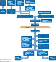
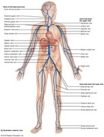
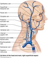
Veins of the Head, Neck, and Brain
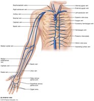
Veins of the Thorax and Right Upper Limb
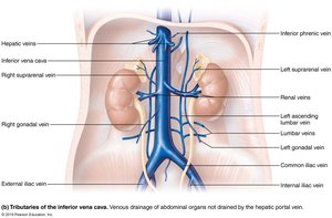
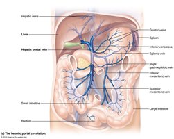
Veins of the Abdomen
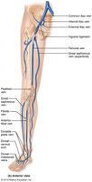
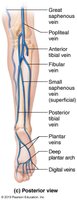
Veins of the Right Lower Limb
Summary Table: Capillary Transport Mechanisms
Route | Example | Type of Molecule |
|---|---|---|
Direct diffusion | O2, CO2 | Lipid-soluble |
Intercellular clefts | Glucose, ions | Water-soluble |
Fenestrations | Small peptides | Water-soluble |
Pinocytotic vesicles | Proteins | Large molecules |
Summary Table: Major Systemic Arteries and Veins
Region | Major Artery | Major Vein |
|---|---|---|
Head/Neck | Carotid arteries | Jugular veins |
Upper Limb | Brachial artery | Basilic/cephalic veins |
Thorax | Thoracic aorta | Intercostal veins |
Abdomen | Abdominal aorta | Inferior vena cava |
Pelvis/Lower Limb | Femoral artery | Femoral vein |
Key Equations
Blood Flow (F): Where is the pressure difference and is resistance.
Mean Arterial Pressure (MAP):
Net Filtration Pressure (NFP): Where HP = hydrostatic pressure, OP = osmotic pressure, c = capillary, if = interstitial fluid.
Clinical Application
Understanding blood vessel structure, function, and regulation is essential for diagnosing and treating cardiovascular disorders, such as hypertension, edema, and vascular occlusions.
