 Indietro
Blood Vessels and Hemodynamics: Structure, Function, and Regulation
Guida di studio - Note intelligenti
 Appunti personalizzati basati sui tuoi materiali, ampliati con definizioni chiave, esempi e contesto.
Blood Vessel Structure and Organization
General Anatomical Organization of Blood Vessels
Blood vessels are tubular structures that transport blood throughout the body. They are organized into arteries, veins, and capillaries, each with distinct structural features and functions. - Arteries carry blood away from the heart under high pressure. - Veins return blood to the heart under lower pressure. - Capillaries facilitate exchange between blood and tissues. 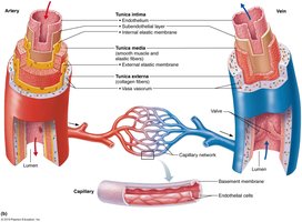
Histological Organization of Blood Vessel Walls
Blood vessel walls are composed of three layers, or tunics: - Tunica intima: Innermost layer, consisting of endothelium (simple squamous epithelium) and basement membrane. - Tunica media: Middle layer, primarily smooth muscle and elastic fibers; responsible for vasoconstriction and vasodilation. - Tunica externa (adventitia): Outermost layer, connective tissue with collagen for protection and reinforcement. 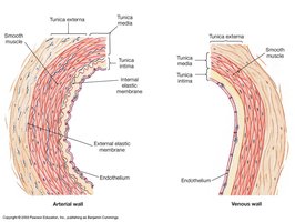
Blood Vessel Wall Structure and Function
- The lumen is the central blood-containing space. - Vasoconstriction (narrowing) and vasodilation (widening) of the lumen are regulated by sympathetic vasomotor nerves acting on the tunica media. - These changes affect blood flow and blood pressure.
Comparison of Arteries and Veins
Arteries and veins differ in wall thickness, lumen size, and structural composition. - Arteries have thicker tunica media and smaller lumens. - Veins have thinner walls, larger lumens, and often contain valves to prevent backflow. 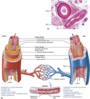
Histological Structure of Blood Vessels
- All vessels contain at least one tunic. - Capillaries consist only of endothelium and basement membrane. 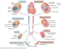
Relative Proportion of Blood Volume Throughout the Cardiovascular System
Blood volume is distributed unevenly: - Systemic veins and venules: 60% - Systemic arteries and arterioles: 15% - Pulmonary blood vessels: 12% - Heart: 8% - Capillaries: 5% 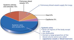
The Arterial System
Types of Arteries
Arteries are classified by size and function: - Elastic arteries: Largest, near the heart (e.g., aorta), with high elastin content for pressure buffering. - Muscular arteries: Medium-sized, deliver blood to organs, thick tunica media for vasomotion. - Arterioles: Smallest, regulate blood flow into capillary beds, major contributors to peripheral resistance. 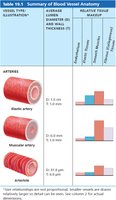
Structural and Functional Differences
- Elastic arteries are highly distensible, allowing them to absorb pressure fluctuations. - Muscular arteries have more smooth muscle, enabling precise control of blood flow. - Arterioles have high resistance, crucial for regulating blood pressure.
Summary Table: Blood Vessel Anatomy
Vessel Type | Diameter | Wall Thickness | Relative Tissue Makeup |
|---|---|---|---|
Elastic artery | 1.0-2.5 cm | 1.0 mm | High elastin, moderate smooth muscle |
Muscular artery | 0.3-1.0 cm | 1.0 mm | High smooth muscle, less elastin |
Arteriole | 10-100 μm | 0.5 μm | Mostly smooth muscle |
Capillaries and Microcirculation
Capillary Beds and Regulation
Capillaries form networks called capillary beds, where exchange occurs. - Blood flow into capillaries is regulated by precapillary sphincters and arterioles. - Only 30-50% of capillaries are open at any time. 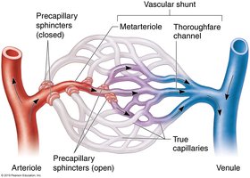
Transition from Arterioles to Capillaries
- Blood leaves arterioles via metarterioles, which branch into capillaries. - Precapillary sphincters control entry into capillaries.
Venous System
- Capillaries unite to form venules, which empty into veins. - Veins return blood to the heart and act as blood reservoirs. 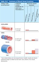
Hemodynamics: Blood Flow, Pressure, and Resistance
Principles of Blood Flow
Hemodynamics describes the physical principles governing blood flow. - Blood pressure: Force exerted by blood on vessel walls (mm Hg). - Blood flow: Volume of blood passing through a tissue per unit time (ml/min). - Perfusion: Blood flow per mass of tissue (ml/min/g). - Resistance: Opposition to flow, mainly due to vessel diameter and blood viscosity. - Key equation: (Flow is proportional to pressure difference divided by resistance)
Velocity of Blood Flow
- Velocity is inversely related to total cross-sectional area. - Arteries: Highest velocity (smallest area) - Capillaries: Lowest velocity (largest area) - Veins: Intermediate velocity
Blood Pressure Changes With Distance From Heart
- Blood pressure is highest and most pulsatile near the heart. - Pressure drops significantly in arterioles and becomes steady in capillaries and veins. 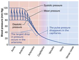
Mean Arterial Pressure (MAP)
- MAP is the average pressure in arteries during one cardiac cycle. - Calculated as: - Also: (Cardiac Output × Total Peripheral Resistance)
Major Factors That Increase MAP
- Increased stroke volume, heart rate, blood vessel diameter, blood viscosity, and vessel length all raise MAP. 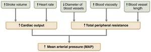
Peripheral Resistance
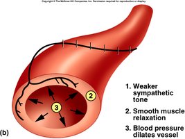
- Resistance is mainly determined by vessel radius, blood viscosity, and vessel length. - Sympathetic stimulation increases wall tension, decreases radius, and raises resistance. 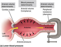 - Decreased sympathetic stimulation relaxes smooth muscle, increases radius, and lowers resistance.
Regulation of Blood Pressure and Flow
Neural, Hormonal, and Local Control
Blood pressure and flow are regulated by: - Neural control: Baroreceptors and chemoreceptors detect changes and signal the medulla. - Hormonal control: Hormones like angiotensin II, epinephrine, and norepinephrine affect vessel tone. - Local control: Metabolic factors and vasoactive chemicals regulate perfusion. 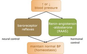
Baroreceptors and Chemoreceptors
- Baroreceptors are located in the carotid sinus and aortic arch; they sense pressure changes. 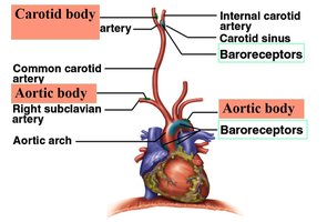 - Chemoreceptors respond to changes in blood chemistry (pH, O2, CO2).
Neural Control: Baroreflex and Chemoreflex
- Baroreflex is a negative feedback mechanism to stabilize blood pressure. 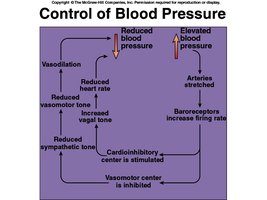 - Chemoreflex adjusts respiration and vasomotion in response to hypoxemia, hypercapnia, and acidosis.
Hormonal Control: Renin-Angiotensin-Aldosterone System (RAAS)
- Angiotensin II is a potent vasoconstrictor, increasing blood pressure. - RAAS is activated by low blood pressure, leading to increased sodium and water retention. 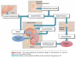
Other Hormones Influencing BP
- Epinephrine and norepinephrine constrict most vessels but dilate those in skeletal and cardiac muscle.
Local Control of BP and Flow
- Metabolic theory: Inadequate perfusion leads to vasodilation. - Vasoactive chemicals (e.g., histamine, bradykinin) stimulate vasomotion. - Angiogenesis: Growth of new vessels in response to tissue needs.
Blood Flow Comparison Between Rest and Exercise
- Exercise increases perfusion to lungs, heart, and skeletal muscles, while decreasing flow to kidneys and digestive tract. 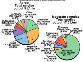
Capillary Structure and Exchange
Types of Capillaries
- Continuous capillaries: Least permeable, most common; found in skin, muscles, lungs, CNS. 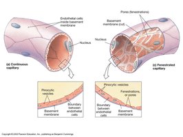 - Fenestrated capillaries: More permeable, found in kidneys, intestines, endocrine glands.
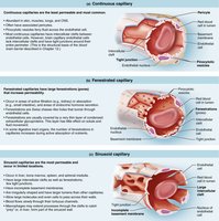 - Sinusoidal capillaries: Most permeable, found in liver, bone marrow, spleen, adrenal medulla.
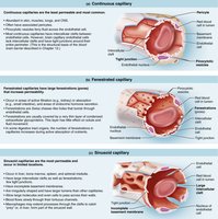
Capillary Exchange Mechanisms
Exchange occurs via: - Intercellular clefts - Fenestrations - Transcytosis (pinocytosis and exocytosis) - Diffusion (lipid-soluble and water-soluble substances) 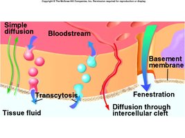
Forces Influencing Capillary Volume and Flow
- Hydrostatic pressure: Pushes fluid out of capillaries. - Colloid osmotic pressure: Pulls fluid into capillaries.
Capillary Filtration and Reabsorption
- Filtration occurs at the arterial end; reabsorption at the venous end. - Net movement depends on the balance of hydrostatic and osmotic pressures. 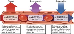
The Venous System
Veins as Capacitance Vessels
- Veins contain up to 65% of blood supply, mainly pooled in the liver, spleen, and skin. 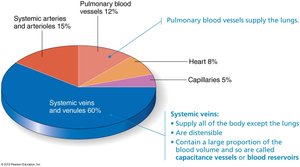
Mechanisms of Venous Return
- Pressure gradient, gravity, skeletal muscle pump, thoracic pump, and cardiac suction all aid venous return.
Other Adaptations of Veins
- Venous sinuses are specialized, flattened veins with thin walls, supported by surrounding tissues.
Major Vessels of the Body
The Great Vessels
- Includes the aorta, pulmonary trunk, superior and inferior vena cava, and pulmonary veins.
The Pulmonary and Systemic Circuits
- Pulmonary circuit: Carries blood from heart to lungs and back. - Systemic circuit: Distributes blood to all body tissues.
Major Arteries and Veins
- Aorta branches into brachiocephalic trunk, left common carotid, and left subclavian arteries. - Major arteries and veins supply and drain the chest, trunk, upper and lower limbs, head, and neck.
Summary Table: Capillaries and Veins
Vessel Type | Diameter | Wall Thickness | Relative Tissue Makeup |
|---|---|---|---|
Capillary | 8-10 μm | 0.5 μm | Endothelium only |
Venule | 20 μm | 1.0 μm | Thin tunica media, endothelium |
Vein | 0.5-3 cm | 0.5 mm | Thin tunica media, thick tunica externa |
Recap
The cardiovascular system's blood vessels are organized into arteries, veins, and capillaries, each with unique structural and functional properties. Blood flow, pressure, and resistance are regulated by neural, hormonal, and local mechanisms, ensuring efficient perfusion and homeostasis. Understanding these principles is essential for comprehending cardiovascular physiology and pathology.
