 Indietro
Blood Vessels: Structure and Function in the Cardiovascular System
Guida di studio - Note intelligenti
 Appunti personalizzati basati sui tuoi materiali, ampliati con definizioni chiave, esempi e contesto.
The Cardiovascular System: Blood Vessels
Overview of Blood Vessels
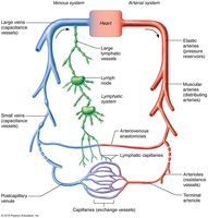
The cardiovascular system consists of a network of blood vessels that transport blood throughout the body, working closely with the lymphatic system to maintain fluid balance and support cellular needs. Blood vessels are classified into arteries, capillaries, and veins, each serving distinct functions in circulation.
Arteries: Carry blood away from the heart.
Capillaries: Facilitate direct exchange of materials with tissue cells.
Veins: Return blood toward the heart.
Structure of Blood Vessel Walls
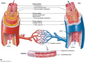
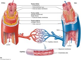
Most blood vessels (except capillaries) have three distinct layers, or tunics, surrounding a central lumen. These layers provide structural integrity and regulate blood flow.
Tunica intima: Innermost layer; consists of endothelium (simple squamous epithelium) that lines the lumen, providing a smooth surface to reduce friction.
Tunica media: Middle layer; composed mainly of smooth muscle and elastin. It is innervated by sympathetic vasomotor fibers, controlling vasoconstriction (decreased lumen diameter) and vasodilation (increased lumen diameter). This layer is crucial for maintaining blood flow and pressure.
Tunica externa: Outermost layer; made of loose collagen fibers that protect, reinforce, and anchor the vessel. It contains nerve fibers and lymphatic vessels, and in large veins, elastic fibers.
Capillary Structure
Capillaries are the smallest blood vessels, consisting of a single layer of endothelial cells. Their thin walls allow efficient diffusion of gases, nutrients, and wastes between blood and interstitial fluid.
Types of Blood Vessels
Arteries
Arteries are classified based on size and function:
Elastic arteries: Thick-walled, large lumen vessels (e.g., aorta); act as pressure reservoirs, expanding and recoiling with each heartbeat.
Muscular arteries: Distribute blood to body organs; have the thickest tunica media with more smooth muscle.
Arterioles: Smallest arteries; control blood flow into capillary beds via vasodilation and vasoconstriction. They are also called resistance arteries.
Capillaries
Capillaries are microscopic vessels that supply nearly every cell. Their primary function is the exchange of gases, nutrients, wastes, and hormones between blood and tissues.
Types of Capillaries
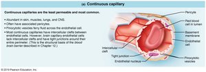
Continuous capillaries: Most common; found in skin, muscles, lungs, and CNS. Endothelial cells are joined by tight junctions with intercellular clefts for passage of fluids and small solutes. In the brain, they form the blood-brain barrier.
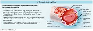
Fenestrated capillaries: Found in areas of active filtration (kidneys), absorption (intestines), or hormone secretion. Endothelial cells have pores (fenestrations) for increased permeability.
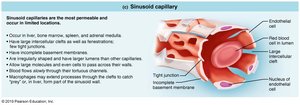
Sinusoidal capillaries: Located in liver, bone marrow, spleen, and adrenal medulla. They have large lumens, fewer tight junctions, and are highly permeable, allowing passage of large molecules and cells. Macrophages are present to capture foreign invaders.
Capillary Beds
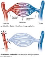
Capillary beds are networks of capillaries between arterioles and venules, facilitating microcirculation. Blood flow through these beds is regulated by the diameter of terminal arterioles and upstream arterioles.
Terminal arteriole: Branches into multiple capillaries for exchange.
Postcapillary venule: Drains capillaries after exchange.
Microcirculation: Blood flow from arteriole to venule through capillary bed.
Veins
Structure and Function
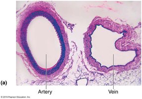
Veins carry blood toward the heart and are formed by the convergence of venules. They have thinner walls and larger lumens compared to arteries, making them effective blood reservoirs.
Venules: Formed by the union of capillaries; highly porous, allowing fluids and white blood cells to enter tissues.
Veins: Have all three tunics, but with a thin tunica media and thick tunica externa. Contain collagen fiber networks and can store up to 65% of blood supply.
Blood Volume Distribution
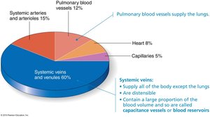
The cardiovascular system distributes blood volume among its components, with veins holding the largest proportion.
Component | Percentage of Blood Volume |
|---|---|
Systemic veins and venules | 60% |
Systemic arteries and arterioles | 15% |
Pulmonary blood vessels | 12% |
Heart | 8% |
Capillaries | 5% |
Adaptations for Venous Return
Because venous blood pressure is lower than arterial, veins have adaptations to ensure blood returns to the heart:
Large-diameter lumens: Reduce resistance.
Venous valves: Prevent backflow; most abundant in veins of limbs.
Venous sinuses: Flattened veins with thin walls, composed only of endothelium (e.g., coronary sinus, dural sinuses).
Clinical Considerations
Varicose Veins and Homeostatic Imbalance
Varicose veins are dilated, painful veins caused by incompetent valves. Factors include heredity, prolonged standing, obesity, pregnancy, and conditions that hinder venous return. Elevated venous pressure can also cause varicosities, such as hemorrhoids.
Example: Prolonged standing or pregnancy can lead to blood pooling in lower limbs, weakening valves.
Hemorrhoids: Varicosities in anal veins due to increased intra-abdominal pressure.
Summary Table: Blood Vessel Types and Features
Type | Main Features | Function |
|---|---|---|
Arteries | Thick walls, high pressure, three tunics | Carry blood away from heart |
Capillaries | Single endothelial layer, small diameter | Exchange of materials |
Veins | Thin walls, large lumens, valves | Return blood to heart, blood reservoir |
Additional info: The notes above expand on brief points with academic context, including definitions, examples, and tables for clarity. Images are included only when directly relevant to the explanation of the adjacent paragraph.
