 Indietro
Bone formation and cartilage.pptx
Guida di studio - Note intelligenti
 Appunti personalizzati basati sui tuoi materiali, ampliati con definizioni chiave, esempi e contesto.
Bone Formation and Ossification
Ossification: Definition and Origins
Ossification is the process of bone formation, beginning in the embryo and continuing throughout life. Most bones originate from the mesoderm, but craniofacial bones develop from neural crest ectoderm. The final step of ossification is calcification, where minerals are deposited in the bone matrix.
Mesenchyme: Embryonic connective tissue from which bones develop.
Two main types of ossification:
Intramembranous ossification (direct from mesenchyme)
Endochondral (intracartilaginous) ossification (via cartilage model)
Intramembranous Ossification
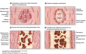
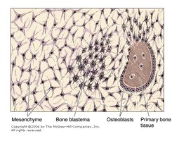
Intramembranous ossification forms flat bones such as those of the skull and clavicle. It involves direct differentiation of mesenchymal cells into bone-forming cells.
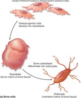
Step 1: Osteoprogenitor cells in mesenchyme differentiate into osteoblasts.
Step 2: Osteoblasts synthesize and partially calcify bone spicules.
Step 3: Bone spicules grow and blood vessels invade the area.
Step 4: Spicules fuse to form a continuous plate of woven bone.
Step 5: Woven bone is remodeled into compact and spongy bone.
Endochondral (Intracartilaginous) Ossification
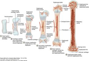
Endochondral ossification forms most bones, including long bones. It involves the replacement of a cartilage model with bone tissue.
Step 1: Fetal hyaline cartilage model develops.
Step 2: Cartilage matrix deteriorates and blood vessels invade.
Step 3: Primary ossification center forms in diaphysis.
Step 4: Secondary ossification centers form in epiphyses.
Step 5: Cartilage is replaced by bone except at articular surfaces and epiphyseal plates.
Bone Growth in Length and Thickness
Longitudinal Growth
Bones grow in length at the epiphyseal plate through endochondral ossification. New cartilage is produced on the epiphyseal side, while bone replaces cartilage on the diaphyseal side.
Epiphyseal plate: Site of active cell division and growth.
Closure: Growth stops when the plate ossifies (epiphyseal closure).
Appositional Growth (Thickness)
Bones grow in thickness by appositional growth, where new bone is added to the periosteum by osteoblasts.
Periosteum: Outer fibrous layer where osteoblasts deposit new bone.
Endosteum: Osteoclasts resorb bone from the inner surface.
Bone Remodeling and Repair
Bone Remodeling
Bone remodeling is a continuous process where old bone is replaced by new bone. It maintains bone strength and mineral homeostasis.
Osteoblasts: Build new bone matrix.
Osteoclasts: Resorb bone matrix.
Remodeling cycle: Activation, resorption, reversal, formation.
Bone Repair
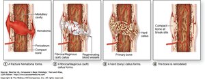
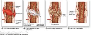
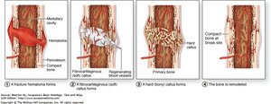
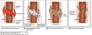
Bone repair after fracture involves several stages, utilizing mechanisms similar to bone remodeling.
Fracture hematoma: Blood vessels break, forming a clot.
Fibrocartilaginous (soft) callus: Macrophages remove debris; fibroblasts and chondroblasts form a soft callus.
Hard (bony) callus: Osteoblasts replace soft callus with trabeculae of primary bone.
Remodeling: Primary bone is remodeled into compact and cancellous bone.
Nutrients and Hormones Regulating Bone Growth
Nutritional Factors
Bone growth and maintenance are highly sensitive to nutritional status, especially during childhood and adolescence.
Calcium: Essential for bone mineralization. Deficiency causes rickets in children and osteomalacia in adults.
Vitamin D: Facilitates calcium absorption.
Other nutrients: Protein, vitamin C, and phosphorus are also important.
Hormonal Regulation
Several hormones regulate bone growth and remodeling:
Parathyroid hormone (PTH): Raises blood calcium by stimulating osteoclast activity.
Calcitonin: Lowers blood calcium by inhibiting osteoclasts.
Growth hormone (GH): Stimulates liver to produce IGF-1, promoting growth at epiphyseal plates.
Sex hormones (androgens and estrogens): Accelerate ossification and closure of epiphyses.
Cartilage: Structure, Function, and Types
Cartilage Overview
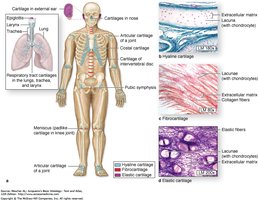
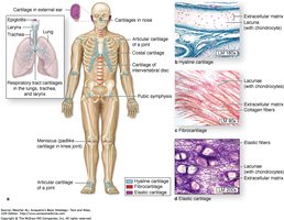
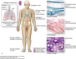
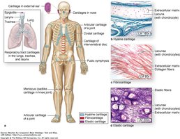
Cartilage is a specialized connective tissue with cells and fibers embedded in a firm matrix. It provides both rigidity and elasticity in various body regions.
Functions:
Serves as the embryonic skeleton.
Allows smooth, friction-free movement of joints.
Acts as a shock absorber.
Types of Cartilage
Cartilage is classified based on the amount and nature of fibers in its matrix:
Hyaline cartilage: Few fibers, glassy appearance.
Fibrocartilage: Abundant collagen fibers, tough and dense.
Elastic cartilage: Contains elastic fibers, flexible.
Hyaline Cartilage
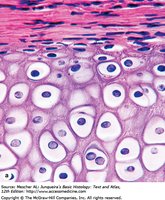
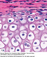
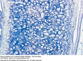
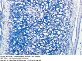
Hyaline cartilage is the most common type, found in articular surfaces, costal cartilages, trachea, nose, and epiphyseal plates.
Structure: Bluish-gray, semitranslucent; perichondrium present except at articular surfaces.
Cells: Chondroblasts and chondrocytes in lacunae, forming isogenous nests.
Matrix: Homogeneous, ground-glass appearance; type II collagen fibers.
Elastic Cartilage
Elastic cartilage is found in the external ear, epiglottis, and certain laryngeal cartilages. It is more flexible than hyaline cartilage.
Structure: Perichondrium present; chondroblasts and chondrocytes are abundant and larger.
Matrix: Numerous branching elastic fibers; territorial and interterritorial matrix.
Fibrocartilage
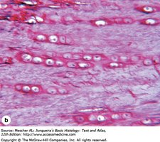
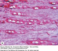
Fibrocartilage is found in intervertebral discs, pubic symphysis, menisci, and articular discs. It is the strongest type of cartilage.
Structure: No perichondrium; bundles of acidophilic type I collagen in matrix.
Cells: Chondrocytes in lacunae arranged in parallel rows.
Function: Shock absorption, weight bearing, contributes to body length.
Summary Table: Types of Cartilage
Type | Fibers | Location | Function |
|---|---|---|---|
Hyaline | Type II collagen | Articular surfaces, trachea, nose, epiphyseal plate | Support, smooth movement |
Elastic | Elastic fibers | External ear, epiglottis | Flexibility |
Fibrocartilage | Type I collagen | Intervertebral discs, pubic symphysis, menisci | Shock absorption, strength |
Additional info:
Bone formation and cartilage topics are directly relevant to Anatomy & Physiology chapters on skeletal tissues, bone growth, and cartilage.
Hormonal and nutritional regulation are essential for understanding bone physiology and pathology.
