 Indietro
Bones and Joints: Structure and Function of the Human Skeleton
Guida di studio - Note intelligenti
 Appunti personalizzati basati sui tuoi materiali, ampliati con definizioni chiave, esempi e contesto.
Bones and Joints: Structure and Function of the Human Skeleton
Overview of the Skeletal System
The human skeleton provides structural support, protection for vital organs, and facilitates movement through its joints. It is composed of 206 bones in adults, organized into the axial and appendicular skeletons.
Axial skeleton: Includes the skull, vertebral column, and thoracic cage.
Appendicular skeleton: Includes the pectoral girdle, pelvic girdle, and limbs.
Vertebral Column
The vertebral column is a flexible structure composed of 26 bones in adults, which include cervical, thoracic, lumbar, sacral, and coccygeal vertebrae. It protects the spinal cord, supports the head, and serves as an anchor for muscles and ligaments.
Cervical (7): Neck region; most flexible.
Thoracic (12): Chest region; ribs attach here.
Lumbar (5): Lower back; largest vertebrae for weight-bearing.
Sacral (5 fused): Forms the sacrum.
Coccygeal (4 fused): Forms the coccyx (tailbone).
Normal Curvatures
The vertebral column has four curvatures that provide resilience and flexibility:
Cervical and lumbar: Concave posteriorly.
Thoracic and sacral: Convex posteriorly.
These curvatures help with weight distribution and shock absorption.
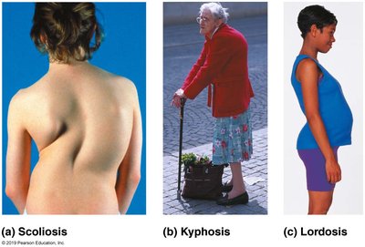
Abnormal Curvatures
Scoliosis: Lateral curvature of the spine.
Kyphosis: Excessive posterior curvature (hunchback).
Lordosis: Excessive anterior curvature (swayback).
Supporting Elements of the Vertebral Column
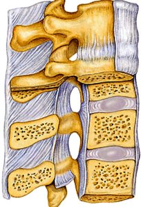
Ligaments and intervertebral discs are essential for stability and flexibility.
Ligaments: Strap-like structures that support the column and prevent excessive movement.
Intervertebral discs: Cushioning pads between vertebrae, composed of a nucleus pulposus (gel-like center) and annulus fibrosus (fibrous ring).
Discs are thickest in the lumbar and cervical regions, contributing to flexibility.
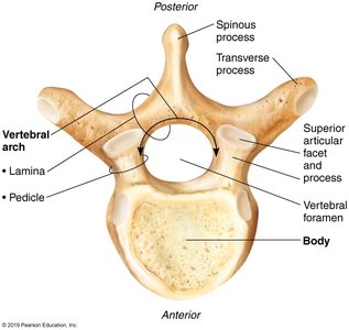
Structure of a Typical Vertebra
Each vertebra consists of a body (weight-bearing), vertebral arch (pedicles and laminae), and several processes for muscle and ligament attachment.
Body: Main weight-bearing region.
Vertebral arch: Forms the vertebral foramen, which houses the spinal cord.
Processes: Spinous, transverse, and articular processes for muscle attachment and articulation.
Regional Differences in Vertebrae
Cervical vertebrae: Small, oval bodies; transverse foramina for vertebral arteries; C1 (atlas) and C2 (axis) have unique features for head movement.
Thoracic vertebrae: Heart-shaped bodies; facets for rib articulation; long spinous processes.
Lumbar vertebrae: Large, kidney-shaped bodies; short, thick processes; triangular vertebral foramen.
Sacrum: Five fused vertebrae; articulates with hip bones.
Coccyx: Four fused vertebrae; attachment for ligaments.
Thoracic Cage
The thoracic cage consists of the thoracic vertebrae, ribs, costal cartilages, and sternum. It protects vital organs and supports the shoulder girdle and upper limbs.
Sternum: Fusion of manubrium, body, and xiphoid process.
Ribs: 12 pairs; true ribs (1-7) attach directly to sternum, false ribs (8-12) do not.
Appendicular Skeleton
The appendicular skeleton includes the pectoral and pelvic girdles, and the limbs. It facilitates movement and supports the body's weight.
Pectoral Girdle
Clavicle: S-shaped bone; braces the shoulder.
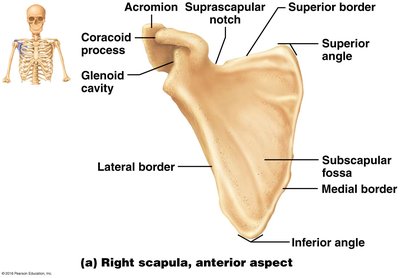
Scapula: Triangular bone; provides attachment points for muscles and articulates with the humerus.
Upper Limb
Humerus: Only bone of the arm; articulates with scapula proximally and radius/ulna distally.
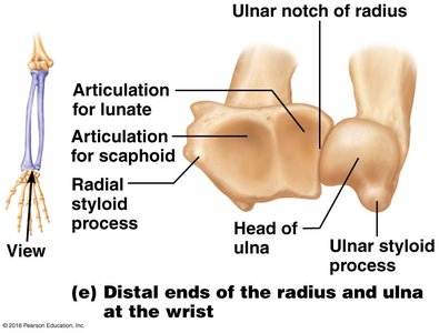
Radius and Ulna: Parallel bones of the forearm; articulate with each other and the humerus.
Carpus (wrist): 8 carpal bones; only scaphoid and lunate articulate with radius.
Metacarpus: 5 metacarpal bones; form the palm.
Phalanges: 14 bones; fingers and thumb (pollex).
Pelvic Girdle
Hip bones (os coxae): Each consists of ilium, ischium, and pubis fused at puberty.
Acetabulum: Socket for the femur; formed by fusion of the three bones.
Lower Limb
Femur: Largest and strongest bone; articulates with pelvis and tibia.
Tibia and Fibula: Parallel bones of the leg; tibia is weight-bearing, fibula is not.
Foot: 26 bones; tarsus (ankle), metatarsus, and phalanges.
Calcaneus: Heel bone; largest tarsal bone.
Hallux: Big toe; has only two phalanges.
Summary Table: Regional Differences in Vertebrae
Region | Body Shape | Key Features |
|---|---|---|
Cervical | Small, oval | Transverse foramina, bifid spinous process |
Thoracic | Heart-shaped | Facets for ribs, long spinous process |
Lumbar | Kidney-shaped | Short, thick processes, triangular foramen |
Sacral | Fused | Articulates with pelvis |
Coccygeal | Fused | Tailbone, ligament attachment |
Key Terms and Definitions
Facet: Smooth surface for articulation.
Foramen: Opening for nerves and blood vessels.
Process: Projection for muscle/ligament attachment.
Condyle: Rounded articular surface.
Epicondyle: Raised area above a condyle.
Equations and Formulas
There are no specific equations for bone anatomy, but the following formula is relevant for calculating bone mass:
Additional info: The notes have been expanded to provide context and definitions for anatomical terms, as well as a summary table for vertebral regional differences. Images included are directly relevant to the anatomical structures discussed.
