 Indietro
Bones and Joints: The Vertebral Column, Thorax, and Appendicular Skeleton
Guida di studio - Note intelligenti
 Appunti personalizzati basati sui tuoi materiali, ampliati con definizioni chiave, esempi e contesto.
Bones & Joints: The Vertebral Column, Thorax, and Appendicular Skeleton
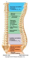
Overview of the Vertebral Column
The vertebral column is a central structure in the human skeleton, providing support, flexibility, and protection for the spinal cord. It consists of 33 bones, with 24 remaining separate for flexibility and 9 fused to form the sacrum and coccyx. The column is approximately 70 cm long in adults of average height.
Regions: 7 cervical, 12 thoracic, 5 lumbar, 5 sacral, 3-4 coccygeal vertebrae
Functions: Weight-bearing, anchor for muscles & ligaments, protection of spinal cord
Normal Curvatures: Cervical & lumbar (concave), thoracic & sacral (convex) for resilience and flexibility
Abnormal Spinal Curvatures
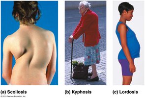
Abnormal curvatures of the spine can affect posture and health. The three main types are:
Scoliosis: Lateral curvature, often in adolescents
Kyphosis: Excessive thoracic curvature, common in elderly
Lordosis: Excessive lumbar curvature, often seen in pregnancy or obesity
Supporting Elements of the Vertebral Column
Ligaments
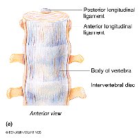
Ligaments are strap-like structures that support the column and keep it upright. The major supporting ligaments are:
Anterior longitudinal ligament: Broad, strongly attached to vertebrae and discs; prevents hyperextension
Posterior longitudinal ligament: Narrow, weaker, attached only to discs; prevents hyperflexion
Intervertebral Discs
Intervertebral discs act as shock absorbers and contribute to 25% of the column's length. Each disc has:
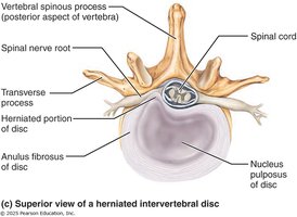
Nucleus pulposus: Gel-like center for elasticity and compressibility
Annulus fibrosus: Tough outer ring that holds vertebrae together and resists tension
Discs are thickest in lumbar and cervical regions for flexibility
Herniated Disc
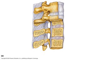
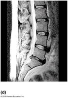
A herniated disc occurs when the nucleus pulposus protrudes through the annulus fibrosus, potentially compressing spinal nerves and causing pain.
Structure of a Typical Vertebra
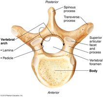
Vertebrae increase in size from cervical to lumbar regions. Each vertebra consists of:
Body: Weight-bearing portion
Vertebral arch: Formed by pedicles and laminae
Vertebral foramen: Encloses the spinal cord
Processes: Spinous, transverse, superior and inferior articular processes for muscle attachment and articulation
Regional Characteristics of Vertebrae
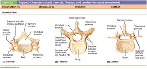
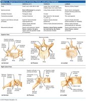
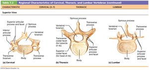
The cervical, thoracic, and lumbar vertebrae have distinct features:
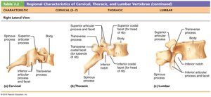
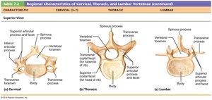
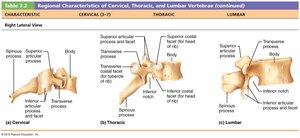
Cervical: Oval body, short bifid spinous process, large vertebral foramen, transverse foramen for vertebral artery
Thoracic: Heart-shaped body, long spinous process, circular vertebral foramen, facets for rib articulation
Lumbar: Kidney-shaped body, flat and short spinous process, triangular vertebral foramen, thick pedicles and laminae
Characteristic | Cervical (C3-C7) | Thoracic | Lumbar |
|---|---|---|---|
Body | Oval | Heart-shaped | Kidney-shaped |
Spinous Process | Short, bifid | Long, points down | Flat, short |
Vertebral Foramen | Large | Circular | Triangular |
Transverse Process | Transverse foramen | Facets for ribs | Thick |
Cervical Vertebrae: Atlas and Axis
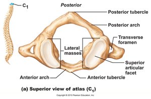
Atlas (C1)
The atlas is the first cervical vertebra, unique in having no body or spinous process. Its superior articular facets articulate with the occipital condyles, allowing the "yes" motion of the head.
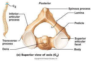
Axis (C2)
The axis is the second cervical vertebra, distinguished by the dens (odontoid process), which acts as a pivot for rotation, allowing the "no" motion of the head.
Thoracic Vertebrae
There are 12 thoracic vertebrae, each attached to ribs. They have heart-shaped bodies, circular vertebral foramina, and long spinous processes. Transverse processes have facets for rib articulation.
Lumbar Vertebrae
The lumbar vertebrae are major weight-bearing bones of the lower back. They have large, kidney-shaped bodies, short and thick pedicles and laminae, and triangular vertebral foramina.
Sacral and Coccygeal Vertebrae
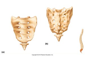
The sacrum is formed by the fusion of five sacral vertebrae during adolescence. It articulates with the fifth lumbar vertebra and the hip bones at the sacroiliac joint. The coccyx (tailbone) consists of 3-4 fused coccygeal vertebrae, serving as an attachment point for pelvic ligaments.
Bony Thorax
Structure and Function
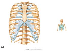
The bony thorax consists of the thoracic vertebrae, ribs, costal cartilages, and sternum. It forms a protective cage around the heart, lungs, and major blood vessels, supports the shoulder girdle and upper limbs, and provides muscle attachment areas.
Sternum
The sternum is the anterior midline bone of the thorax, formed by the fusion of the manubrium, body, and xiphoid process. Key landmarks include:
Jugular notch: Palpable indentation
Sternal angle: Cartilaginous hinge between manubrium and body
Xiphisternal joint: Fusion of sternal body and xiphoid process
Ribs
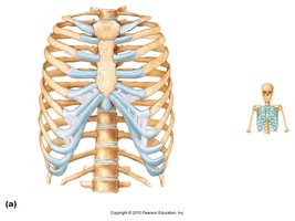
There are 12 pairs of ribs, all attaching posteriorly to the vertebral column. Ribs curve inferiorly and anteriorly. Classification:
True ribs (1-7): Attach directly to sternum
False ribs (8-10): Attach indirectly via costal cartilages
Floating ribs (11-12): Not attached anteriorly
Structure of a Typical Rib
A typical rib is a bowed, flat bone with a shaft, head, neck, and tubercle. The head has two facets for articulation with thoracic vertebrae, and the tubercle articulates with the transverse process of the same-numbered vertebra.
Appendicular Skeleton
Pectoral Girdle
The pectoral girdle consists of the clavicles and scapulae, forming a nearly complete circle around the upper trunk. It provides attachment points for muscles that move the upper limbs and is highly mobile.
Clavicles: S-shaped, act as braces to push arms laterally
Scapulae: Thin, triangular, flat bones located dorsally between ribs 2 and 7
Upper Limb
The upper limb contains 30 bones, including the arm (humerus), forearm (radius and ulna), wrist (carpals), and hand (metacarpals and phalanges).
Humerus: Longest bone of the upper limb, articulates with scapula proximally and radius/ulna distally
Ulna: Slightly longer than radius, forms elbow joint
Radius: Contributes to wrist joint
Carpus: 8 carpal bones; only scaphoid and lunate articulate with radius
Metacarpus: 5 long bones, numbered 1-5 from thumb to little finger
Phalanges: 3 per finger, 2 in thumb (pollex)
Pelvic Girdle
The pelvic girdle attaches the lower limbs, transmits weight, and supports pelvic organs. It consists of two hip bones (os coxa), each formed by the fusion of the ilium, ischium, and pubis at puberty. The acetabulum is the socket for the hip joint.
Lower Limb
The lower limb includes the thigh (femur), leg (tibia and fibula), and foot (tarsals, metatarsals, phalanges).
Femur: Largest, longest, and strongest bone; articulates with tibia at knee
Tibia: Weight-bearing bone of the leg
Fibula: Lateral bone, not part of knee joint
Foot: 26 bones; tarsus (ankle), metatarsus, and phalanges
Tarsus: 7 tarsal bones; calcaneus (heel) and talus (ankle joint)
Phalanges: Big toe (hallux) has 2, other toes have 3 each
Summary Table: Vertebral Column Regions
Region | Number of Vertebrae | Key Features |
|---|---|---|
Cervical | 7 | Transverse foramen, bifid spinous process |
Thoracic | 12 | Facets for ribs, long spinous process |
Lumbar | 5 | Large body, short spinous process |
Sacral | 5 (fused) | Articulates with pelvis |
Coccygeal | 3-4 (fused) | Tailbone |
Key Terms and Definitions
Vertebral foramen: Opening for the spinal cord
Intervertebral disc: Cushioning structure between vertebrae
Facet: Smooth surface for articulation
Acetabulum: Socket of the hip joint
Pollex: Thumb
Hallux: Big toe
Important Equations
There are no specific equations for bone anatomy, but the following formula is relevant for calculating the total number of vertebrae:
Additional info: Academic context and expanded explanations were added to ensure completeness and clarity for exam preparation.
