 Indietro
Bones and Skeletal Tissue: Structure, Function, and Disorders
Guida di studio - Note intelligenti
 Appunti personalizzati basati sui tuoi materiali, ampliati con definizioni chiave, esempi e contesto.
Bones and Skeletal Tissue
Overview of Skeletal Cartilages
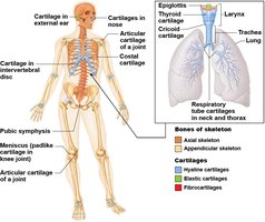
The human skeleton initially consists of cartilage, which is gradually replaced by bone except in areas requiring flexibility. Cartilage is a resilient and flexible connective tissue found in various parts of the body.
Hyaline cartilage: Provides support, flexibility, and resilience; most abundant; found in articular (joints), costal (ribs), respiratory (larynx), and nasal cartilage.
Elastic cartilage: Similar to hyaline but contains elastic fibers; found in external ear and epiglottis.
Fibrocartilage: Contains thick collagen fibers; great tensile strength; found in menisci of knee and intervertebral discs.
Functions of Bones
Bones serve several essential functions in the human body, contributing to both structure and physiology.
Support: Provides framework for the body and supports soft organs.
Protection: Shields vital organs such as the brain, spinal cord, and thoracic organs.
Movement: Acts as levers for muscles, enabling movement.
Mineral and growth factor storage: Reservoir for calcium, phosphorus, and growth factors.
Blood cell formation: Hematopoiesis occurs in red marrow cavities.
Triglyceride storage: Fat stored in bone cavities serves as an energy source.
Hormone production: Osteocalcin regulates insulin secretion, glucose levels, and metabolism.
Classification of Bones
The human skeleton contains 206 named bones, classified by location and shape.
Axial skeleton: Long axis of body; includes skull, vertebral column, and rib cage.
Appendicular skeleton: Bones of limbs and girdles attaching limbs to axial skeleton.
Bones are also classified by shape:
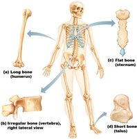
Long bones: Longer than wide; found in limbs.
Short bones: Cube-shaped; found in wrist and ankle; sesamoid bones form within tendons (e.g., patella).
Flat bones: Thin, flat, slightly curved; includes sternum, scapulae, ribs, and most skull bones.
Irregular bones: Complicated shapes; includes vertebrae and hip bones.
Bone Structure
Gross Anatomy of Bone
Bones are organs composed of several tissue types, including osseous tissue, nervous tissue, cartilage, fibrous connective tissue, muscle cells, and epithelial cells in blood vessels. Bone structure is studied at three levels: gross, microscopic, and chemical.
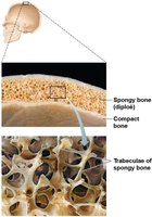
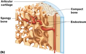
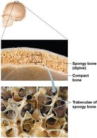
Compact bone: Dense outer layer; appears smooth and solid.
Spongy bone: Honeycomb structure of trabeculae; spaces filled with red or yellow marrow.
Structure of Long Bones
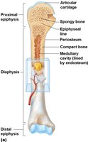
Long bones have a distinct anatomy, including a shaft (diaphysis), ends (epiphyses), and membranes.
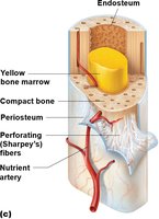
Diaphysis: Tubular shaft; compact bone surrounds medullary cavity filled with yellow marrow in adults.
Epiphyses: Bone ends; compact bone externally, spongy bone internally; articular cartilage covers joint surfaces.
Epiphyseal line: Remnant of childhood epiphyseal plate where bone growth occurs.
Hematopoietic Tissue in Bones
Red marrow is found within trabecular cavities of spongy bone and diploë of flat bones. In newborns, all spongy bone and medullary cavities contain red marrow; in adults, red marrow is mainly in flat bones and some irregular bones. Yellow marrow can convert to red if needed.
Microscopic Anatomy of Bone
Bone Cells
Bone tissue contains five major cell types, each specialized for specific functions.
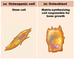
Osteogenic cells: Stem cells in periosteum and endosteum; differentiate into osteoblasts or bone-lining cells.
Osteoblasts: Bone-forming cells; secrete osteoid (collagen and calcium-binding proteins).
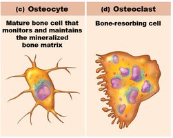
Osteocytes: Mature bone cells in lacunae; maintain matrix and act as stress sensors.
Bone-lining cells: Flat cells on bone surfaces; help maintain matrix.
Osteoclasts: Bone-resorbing cells; break down bone matrix.
Structure of Compact Bone
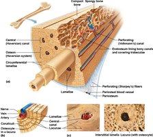
Compact bone, also called lamellar bone, consists of osteons, canals, canaliculi, and lamellae.
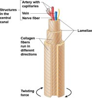
Osteon (Haversian system): Structural unit; elongated cylinder with concentric lamellae.
Lamellae: Rings of bone matrix; collagen fibers run in different directions to resist twisting.
Central (Haversian) canal: Contains blood vessels and nerves.
Perforating (Volkmann’s) canals: Connect blood vessels and nerves of periosteum, medullary cavity, and central canal.
Lacunae: Small cavities containing osteocytes.
Canaliculi: Hairlike canals connecting lacunae; allow communication and nutrient/waste exchange.
Structure of Spongy Bone
Spongy bone is organized along lines of stress, with trabeculae conferring strength. Trabeculae contain irregularly arranged lamellae and osteocytes interconnected by canaliculi. Capillaries in endosteum supply nutrients.
Chemical Composition of Bone
Organic Components
Organic components include cells (osteogenic, osteoblasts, osteocytes, bone-lining cells, osteoclasts) and osteoid (ground substance and collagen fibers). Osteoid contributes to tensile strength and flexibility.
Inorganic Components
Inorganic components are mainly hydroxyapatites (mineral salts), which make up 65% of bone mass. These tiny calcium phosphate crystals provide hardness and resistance to compression.
Bone is half as strong as steel in resisting compression and as strong as steel in resisting tension.
Mineral composition allows bone to last long after death and provides information about ancient populations.
Formation of the Bony Skeleton
Endochondral Ossification
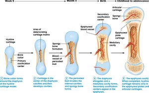
Endochondral ossification is the process by which bone forms by replacing hyaline cartilage. Most bones of the skeleton are formed this way.
Primary ossification center forms in the diaphysis.
Cartilage in the center of the diaphysis calcifies and develops cavities.
Periosteal bud invades cavities, forming spongy bone.
Diaphysis elongates and medullary cavity forms.
Epiphyses ossify, leaving articular cartilage and epiphyseal plate.
Intramembranous Ossification
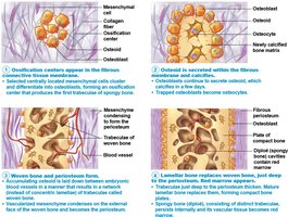
Intramembranous ossification begins within fibrous connective tissue membranes formed by mesenchymal cells. It forms flat bones such as the frontal, parietal, occipital, temporal, and clavicle bones.
Ossification centers appear in fibrous connective tissue.
Osteoid is secreted and calcifies.
Woven bone and periosteum form.
Lamellar bone replaces woven bone; red marrow appears.
Bone Disorders
Osteomalacia and Rickets
Osteomalacia is characterized by poorly mineralized bones, resulting in soft, weak bones and pain upon bearing weight. Rickets is the childhood form, causing bowed legs and bone deformities due to vitamin D deficiency or insufficient dietary calcium.
Osteoporosis
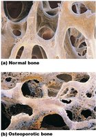
Osteoporosis is a group of diseases where bone resorption exceeds bone deposit, leading to decreased bone mass. The spongy bone of the spine and neck of femur is most susceptible, resulting in common vertebral and hip fractures.
Paget’s Disease
Paget’s disease involves abnormal bone remodeling, resulting in bones that are enlarged, deformed, and structurally unsound.
Summary Table: Types of Bone Cells
Cell Type | Function |
|---|---|
Osteogenic cell | Stem cell; differentiates into osteoblasts |
Osteoblast | Matrix-synthesizing cell; responsible for bone growth |
Osteocyte | Mature bone cell; maintains bone matrix |
Bone-lining cell | Flat cell; maintains bone matrix |
Osteoclast | Bone-resorbing cell; breaks down bone matrix |
Summary Table: Classification of Bones by Shape
Shape | Example |
|---|---|
Long bone | Humerus |
Short bone | Talus |
Flat bone | Sternum |
Irregular bone | Vertebra |
