 Indietro
Bones and Skeletal Tissues: Development, Growth, Remodeling, Repair, and Disorders
Guida di studio - Note intelligenti
 Appunti personalizzati basati sui tuoi materiali, ampliati con definizioni chiave, esempi e contesto.
Bones and Skeletal Tissues
Bone Development (Ossification)
Ossification, also known as osteogenesis, is the process by which bone tissue forms. This process begins in the second month of embryonic development and continues throughout life as bones grow, remodel, and repair.
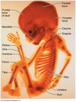
Formation of the Bony Skeleton: Up to week 8, the fetal skeleton consists of fibrous membranes and hyaline cartilage, which are gradually replaced by bone tissue.
Endochondral Ossification: Bone forms by replacing hyaline cartilage. Most bones below the skull (except clavicles) are formed this way.
Intramembranous Ossification: Bone develops directly from fibrous membranes, forming membrane bones such as the frontal, parietal, occipital, temporal, and clavicle bones.
Endochondral Ossification
This process forms most bones of the body and involves several key steps:
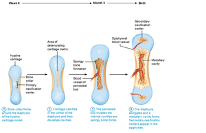
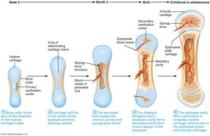
Bone collar forms around the diaphysis of the cartilage model.
Central cartilage in diaphysis calcifies and develops cavities.
Periosteal bud invades cavities, forming spongy bone.
Diaphysis elongates, medullary cavity forms, and secondary ossification centers appear in epiphyses.
Epiphyses ossify; hyaline cartilage remains only in epiphyseal plates and articular cartilages.
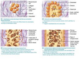
Intramembranous Ossification
Intramembranous ossification forms flat bones of the skull and clavicle. The process involves:
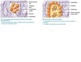
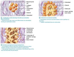
Ossification centers form as mesenchymal cells cluster and become osteoblasts.
Osteoid is secreted and calcified.
Woven bone forms around blood vessels, resulting in trabeculae.
Outer layer forms periosteum; lamellar bone replaces woven bone, and red marrow appears.
Postnatal Bone Growth
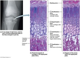
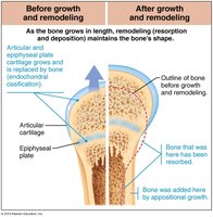
After birth, bones grow in length and width. Long bones grow lengthwise by interstitial growth at the epiphyseal plate, while appositional growth increases bone thickness.
Epiphyseal Plate: Maintains constant thickness; consists of five zones: resting, proliferation, hypertrophic, calcification, and ossification.
Growth in Length: New cartilage is produced on the epiphyseal side, while bone replaces cartilage on the diaphyseal side.
Growth in Width: Osteoblasts add bone matrix to the external surface, while osteoclasts remove bone from the internal surface.
Hormonal Regulation of Bone Growth
Bone growth is regulated by several hormones:
Growth Hormone: Stimulates epiphyseal plate activity in childhood.
Thyroid Hormone: Modulates growth hormone activity for proper proportions.
Sex Hormones: Testosterone and estrogens promote growth spurts and induce epiphyseal plate closure.
Bone Remodeling
Bone remodeling is a lifelong process involving bone deposit and resorption. About 5–7% of bone mass is recycled weekly. Remodeling occurs at periosteum and endosteum surfaces, coordinated by osteoblasts and osteoclasts.
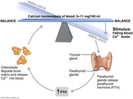
Bone Resorption: Osteoclasts break down bone matrix, releasing calcium into the blood.
Bone Deposit: Osteoblasts deposit new bone matrix, which is then mineralized.
Hormonal Control of Remodeling
Remodeling is regulated by hormones:
Parathyroid Hormone (PTH): Increases bone resorption when blood calcium is low.
Calcitonin: Lowers blood calcium levels at high doses.
Leptin and Serotonin: Influence bone density and turnover.
Mechanical Stress and Wolf’s Law
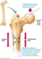
Bones adapt to mechanical stress. Wolf’s law states that bones grow or remodel in response to the demands placed on them. Stress causes electrical signals that stimulate remodeling.
Bone Repair
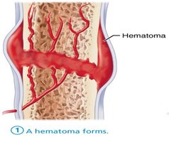
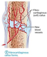
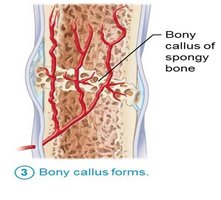
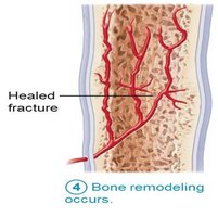
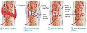
Fractures are breaks in bone, classified by position, completeness, and whether skin is penetrated. Repair involves four stages:
Hematoma formation
Fibrocartilaginous callus formation
Bony callus formation
Bone remodeling
Common Types of Fractures
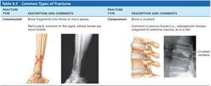
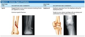
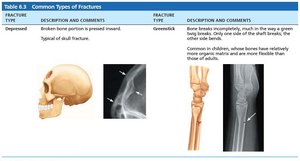
Fractures are classified by their characteristics. The following table summarizes common types:
Fracture Type | Description and Comments |
|---|---|
Comminuted | Bone fragments into three or more pieces. Common in aged bones. |
Compression | Bone is crushed. Common in porous bones. |
Spiral | Ragged break due to twisting forces. Common sports fracture. |
Epiphyseal | Epiphysis separates from diaphysis along epiphyseal plate. |
Depressed | Broken bone portion is pressed inward. Typical of skull fracture. |
Greenstick | Bone breaks incompletely, common in children. |
Bone Disorders
Imbalances between bone deposit and resorption cause bone diseases:
Osteomalacia and Rickets: Poor mineralization leads to soft, weak bones. Rickets affects children, causing bone deformities. Causes include vitamin D deficiency and insufficient calcium.
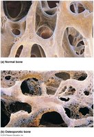
Osteoporosis: Bone resorption exceeds deposit, leading to decreased bone mass and increased fracture risk. Most common in aged, postmenopausal women.
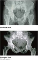
Paget’s Disease: Excessive and disorganized bone remodeling results in weak, deformed bones. Most often affects spine, pelvis, femur, and skull.
Developmental Aspects and Age-Related Changes
Bone formation exceeds resorption in children and adolescents, is balanced in young adults, and resorption exceeds formation in older adults. Bone mass and healing ability decrease with age, especially in females and whites.
Summary Table: Bone Disorders
Disease | Key Features | Causes | Treatment |
|---|---|---|---|
Osteomalacia/Rickets | Soft, weak bones; deformities | Vitamin D deficiency, low calcium | Vitamin D, calcium supplements |
Osteoporosis | Low bone mass, fractures | Aging, menopause, genetics, lifestyle | Calcium, vitamin D, exercise, medications |
Paget’s Disease | Disorganized bone, deformities | Unknown, possibly viral | Calcitonin, bisphosphonates |
Key Terms and Concepts
Ossification: Formation of bone tissue.
Epiphyseal Plate: Growth plate in long bones.
Osteoblast: Bone-forming cell.
Osteoclast: Bone-resorbing cell.
Trabeculae: Lattice-like structure in spongy bone.
Wolf’s Law: Bone adapts to mechanical stress.
Important Equations
Calcium homeostasis is maintained within a narrow range:
Bone remodeling is regulated by the balance between bone deposit and resorption:
Additional info: Bone mass and density are influenced by genetics, nutrition, hormonal status, and physical activity. Early adulthood is critical for maximizing bone mass to buffer against age-related bone loss.
