 Indietro
Bones and Skeletal Tissues: Structure, Function, and Development
Guida di studio - Note intelligenti
 Appunti personalizzati basati sui tuoi materiali, ampliati con definizioni chiave, esempi e contesto.
Bones and Skeletal Tissues
Introduction
The skeletal system is composed of bones and cartilages, providing structural support, protection, movement, storage, and blood cell formation. Understanding the anatomy and physiology of bones and skeletal tissues is fundamental for students of Anatomy & Physiology.
Skeletal Cartilages
Structure of Skeletal Cartilages
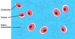
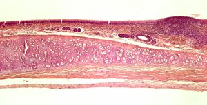
Cartilage is a firm but flexible connective tissue. It consists of chondrocytes (cartilage cells) located in lacunae within a jelly-like extracellular matrix. The perichondrium is a layer of dense irregular connective tissue that surrounds most cartilages, supplying nutrients and providing structural support.
Chondrocytes: Cells responsible for maintaining the cartilage matrix.
Lacunae: Small cavities housing chondrocytes.
Extracellular Matrix: Gel-like substance rich in fibers and ground substance.
Perichondrium: Surrounds cartilage, aids in growth and repair.
Types of Cartilage
There are three main types of cartilage, each with distinct structural and functional properties:
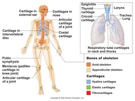
Hyaline Cartilage: Most abundant; contains only collagen fibers. Found in articular surfaces of joints, costal cartilages, respiratory structures, and nose.
Elastic Cartilage: Contains both collagen and elastic fibers. Found in external ear and epiglottis.
Fibrocartilage: Contains thick collagen fibers. Found in menisci of knee, pubic symphysis, and intervertebral discs.
Growth of Cartilage
Cartilage grows by two mechanisms:
Appositional Growth: New matrix is laid down on the surface of cartilage.
Interstitial Growth: Chondrocytes divide within lacunae and secrete new matrix.
Classification and Gross Anatomy of Bones
Functions of Bones
Bones serve several essential functions:
Support: Provide a rigid framework for the body.
Protection: Shield internal organs.
Movement: Act as levers with skeletal muscles.
Storage: Store minerals (calcium, phosphorus) and fat (yellow bone marrow).
Blood Cell Formation: Occurs in red bone marrow.
Classification of Bones
Bones are classified by location and shape:
Axial Skeleton: Skull, vertebral column, thoracic cage.
Appendicular Skeleton: Pectoral girdles, upper limbs, pelvic girdles, lower limbs.
Long Bones: Longer than wide (e.g., humerus).
Short Bones: Cube-shaped (e.g., wrist, ankle bones).
Flat Bones: Thin, flat, slightly curved (e.g., sternum, ribs, cranial bones).
Irregular Bones: Complicated shapes (e.g., vertebrae, hip bones).
Compact and Spongy Bone
Bones contain two types of tissue:
Compact Bone: Dense, hard outer layer.
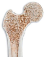
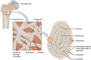
Spongy Bone: Porous interior, composed of trabeculae (network of rods and plates).
Structure of a Typical Long Bone
Long bones have a distinct anatomy:
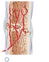
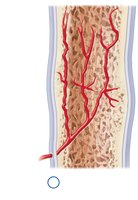
Diaphysis: Tubular shaft, compact bone surrounding medullary cavity (stores yellow bone marrow).
Epiphyses: Wider ends, primarily spongy bone, covered by articular cartilage.
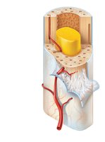
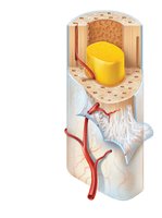
Membranes of Long Bones
Periosteum: Double-layered membrane covering outer surface; outer fibrous layer (dense irregular connective tissue), inner osteogenic layer (stem cells for growth and repair).
Endosteum: Delicate membrane lining inner surfaces (medullary cavity, trabeculae, canals); contains stem cells.
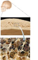
Structure of Flat Bones
Flat bones consist of a layer of spongy bone (diploë) between two layers of compact bone. Red bone marrow fills spaces between trabeculae.
Bone Marrow
Red Bone Marrow: Site of blood cell production; in infants, found in medullary cavities and all spongy bone; in adults, mainly in axial skeleton.
Yellow Bone Marrow: Fat storage; replaces red marrow in medullary cavities of adults.
Microscopic Anatomy of Bone
Types of Bone Cells
Bone contains four primary cell types:
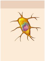
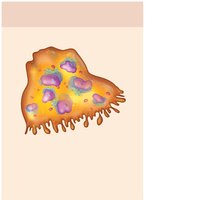
Osteogenic Cells: Stem cells in periosteum and endosteum; some become osteoblasts.
Osteoblasts: Bone-forming cells; secrete unmineralized bone matrix (osteoid).
Osteocytes: Mature bone cells; monitor and maintain bone matrix; located in lacunae.
Osteoclasts: Bone-resorbing cells; derived from white blood cells.
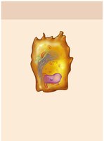
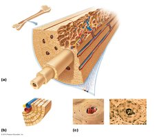
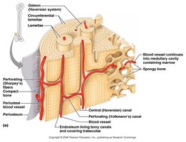
Microscopic Anatomy of Compact Bone
The structural unit of compact bone is the osteon (Haversian system), an elongated cylinder running parallel to the bone's long axis. Osteons resist stress and bending.
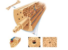
Central Canal: Contains blood vessels and nerve fibers.
Lamellae: Concentric rings of bone matrix surrounding the central canal.
Lacunae: Small cavities containing osteocytes.
Canaliculi: Tiny canals connecting osteocytes and central canal, allowing nutrient and waste exchange.
Perforating Canals: Run perpendicular to the bone's axis, connecting central canals and periosteum.
Microscopic Anatomy of Spongy Bone
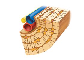
Spongy bone lacks osteons. Its trabeculae contain osteocytes in lacunae, interconnected by canaliculi.
Chemical Composition of Bone
Bones are composed of both organic and inorganic components:
Organic (35%): Collagen, ground substance, living cells; provides flexibility and tensile strength.
Inorganic (65%): Mineral salts (mainly calcium phosphate); provides hardness and resistance to compression.
Bone Development
Bone Formation (Ossification)
Ossification (osteogenesis) is the process of bone formation, beginning in the second month of development and continuing throughout life.
Formation of Bony Skeleton: Two pathways: intramembranous and endochondral ossification.
Postnatal Bone Growth: Continues until early adulthood.
Bone Remodeling and Repair: Lifelong process.
Intramembranous Ossification
Bone develops within a fibrous membrane, forming flat bones of the skull, most facial bones, and clavicles.
Ossification centers form.
Osteoid is secreted and calcifies.
Immature spongy bone and periosteum form.
Compact bone develops and red bone marrow appears.
Endochondral Ossification
Bone develops by replacing hyaline cartilage. Forms all bones below the head except clavicles.
Mesenchymal cells differentiate into chondroblasts, producing a hyaline cartilage model.
Ossification begins in the primary ossification center in the shaft.
Perichondrium transforms into periosteum; osteoblasts appear.
Secondary ossification centers appear in epiphyses after birth.
Growth and Remodeling of Bones
Growth in Length of Long Bones
Long bones grow in length at the epiphyseal plates:
Cartilage forms on the epiphyseal side.
Cartilage is ossified on the diaphyseal side.
Diaphysis elongates.
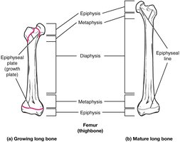
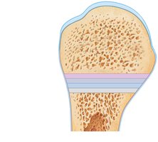
Epiphyseal plate has five zones: resting, proliferation, hypertrophic, calcification, ossification.
Epiphyseal plate closure occurs when chondrocytes stop dividing and cartilage is replaced by bone, forming the epiphyseal line.
Growth in Width (Diameter)
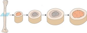
Bones increase in diameter through appositional growth:
Osteoclasts remove old bone along the medullary cavity.
Osteoblasts secrete new bone beneath the periosteum.
Bone Remodeling
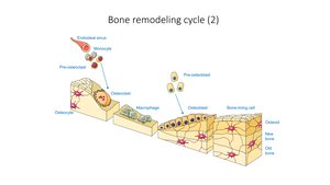
Bone remodeling is a lifelong process where old bone tissue is replaced by new bone, involving bone resorption (osteoclasts) and bone deposit (osteoblasts).
Control of Remodeling
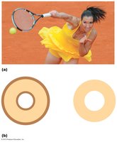
Mechanical Stress: Bones grow or remodel in response to demands placed on them.
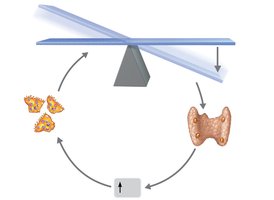
Calcium Homeostasis: Maintained by parathyroid hormone (PTH) and calcitonin.
Bone Repair and Disorders
Bone Repair
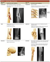
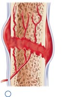
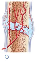
Fracture repair involves four stages:
Hematoma forms (days 1–7): Blood clots at fracture site.
Fibrocartilaginous callus forms (weeks 1–3): Collagen and fibrocartilage replace clot.
Bony callus forms (weeks 3–8): Soft callus replaced by spongy bone.
Bone remodeling occurs (months to years): Osteoclasts remove excess bone, osteoblasts deposit new compact bone.
Bone Disorders
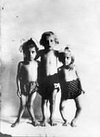
Osteomalacia and Rickets: Inadequate mineralization due to vitamin D or calcium deficiency; leads to weak, softened bones. Osteomalacia affects adults, rickets affects children.
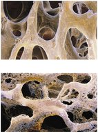
Osteoporosis: Bone resorption outpaces bone deposit; bone mass declines, matrix remains normal. Influenced by hormonal changes, especially loss of estrogen after menopause.
Type of Bone Cell | Function |
|---|---|
Osteogenic Cell | Stem cell for bone growth and repair |
Osteoblast | Bone-forming cell; secretes osteoid |
Osteocyte | Mature bone cell; maintains bone matrix |
Osteoclast | Bone-resorbing cell; breaks down bone |
Type of Cartilage | Main Fibers | Locations |
|---|---|---|
Hyaline | Collagen | Joints, ribs, nose, larynx, trachea |
Elastic | Collagen & Elastic | External ear, epiglottis |
Fibrocartilage | Thick Collagen | Knee menisci, pubic symphysis, intervertebral discs |
Key Equations:
Calcium homeostasis:
