 Indietro
Bones and Skeletal Tissues: Structure, Function, and Development
Guida di studio - Note intelligenti
 Appunti personalizzati basati sui tuoi materiali, ampliati con definizioni chiave, esempi e contesto.
Bones and Skeletal Tissues
Overview of Skeletal Cartilage
The human skeleton initially consists of cartilage, which is gradually replaced by bone except in areas requiring flexibility. Skeletal cartilage is a resilient tissue composed mainly of water, lacking blood vessels and nerves. The perichondrium, a dense connective tissue layer, surrounds cartilage and provides nutrients via blood vessels.
Chondrocytes: Cells encased in lacunae within a jelly-like extracellular matrix.
Types of Cartilage:
Hyaline cartilage: Provides support, flexibility, and resilience; found in joints, ribs, larynx, and nose.
Elastic cartilage: Contains elastic fibers; found in the external ear and epiglottis.
Fibrocartilage: Contains thick collagen fibers; found in menisci of the knee and vertebral discs.
Growth of Cartilage
Cartilage grows by two mechanisms:
Appositional growth: Cartilage-forming cells in the perichondrium secrete matrix on the surface.
Interstitial growth: Chondrocytes within lacunae divide and secrete new matrix internally.
Calcification: Occurs during bone growth in youth and old age, but hardened cartilage is not bone.
Functions of Bones
Bones serve seven essential functions:
Support: Framework for the body and soft organs.
Protection: Shields the brain, spinal cord, and vital organs.
Movement: Acts as levers for muscle action.
Mineral and growth factor storage: Reservoir for calcium, phosphorus, and growth factors.
Blood cell formation: Hematopoiesis occurs in red marrow cavities.
Triglyceride storage: Fat stored in bone cavities for energy.
Hormone production: Osteocalcin regulates insulin secretion and metabolism.
Classification of Bones
The human skeleton contains 206 named bones, divided into two groups:
Axial skeleton: Skull, vertebral column, rib cage.
Appendicular skeleton: Limbs and girdles attaching limbs to the axial skeleton.
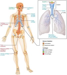
Classification by Shape
Long bones: Longer than wide; found in limbs.
Short bones: Cube-shaped; found in wrists and ankles. Sesamoid bones form within tendons (e.g., patella).
Flat bones: Thin, flat, slightly curved; found in the sternum, scapulae, ribs, and skull.
Irregular bones: Complex shapes; found in vertebrae and hip bones.
Bone Structure
Bones are organs containing various tissues: bone (osseous), nervous, cartilage, fibrous connective, muscle, and epithelial cells. Structure is studied at three levels: gross, microscopic, and chemical.
Gross Anatomy
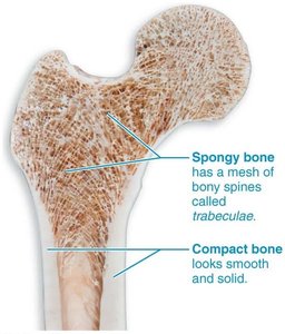
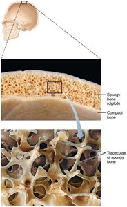
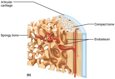
Compact bone: Dense outer layer, smooth and solid.
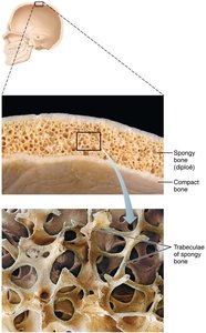
Spongy bone: Honeycomb of trabeculae, with spaces filled by red or yellow marrow.
Structure of Short, Irregular, and Flat Bones
Thin plates of spongy bone (diploe) covered by compact bone.
Periosteum covers outside; endosteum covers inside.
Bone marrow scattered throughout spongy bone; no defined cavity.
Hyaline cartilage covers areas involved in movable joints.
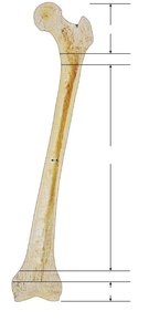
Structure of Long Bones
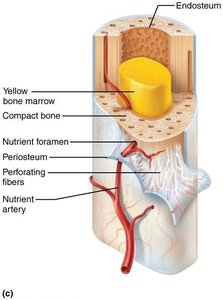
Diaphysis: Tubular shaft, compact bone surrounds medullary cavity (yellow marrow in adults).
Epiphyses: Ends of long bones, compact bone externally, spongy bone internally; articular cartilage covers joint surfaces.
Epiphyseal line: Remnant of childhood epiphyseal plate.
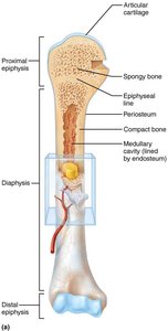
Membranes
Periosteum: Double-layered membrane covering external surfaces except joints; fibrous layer (dense irregular connective tissue), osteogenic layer (stem cells).
Endosteum: Delicate membrane covering internal bone surfaces, trabeculae, and canals; contains osteogenic cells.
Hematopoietic Tissue in Bones
Red marrow: Found in trabecular cavities of spongy bone and diploë of flat bones; in adults, mainly in femur, humerus, hip, and sternum.
Yellow marrow: Can convert to red marrow if needed.
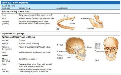
Bone Markings
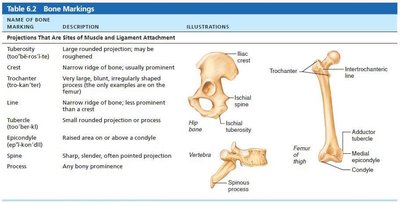
Bone markings are sites for muscle, ligament, and tendon attachment, joint formation, or passageways for blood vessels and nerves. They are classified as projections, depressions, or openings.
Name of Bone Marking | Description | Illustration |
|---|---|---|
Tuberosity | Large rounded projection | Hip bone |
Crest | Narrow ridge of bone | Hip bone |
Trochanter | Very large, irregularly shaped process | Femur |
Line | Narrow ridge of bone | Femur |
Tubercle | Small rounded projection | Femur |
Epicondyle | Raised area above a condyle | Femur |
Spine | Sharp, slender projection | Vertebra |
Process | Any bony prominence | Spinous process |
Name of Bone Marking | Description | Illustration |
|---|---|---|
Facet | Smooth, nearly flat articular surface | Rib |
Condyle | Rounded articular projection | Mandible |
Groove | Furrow | Skull |
Foramen | Round or oval opening | Skull |
Notch | Indentation at edge of structure | Skull |
Meatus | Canal-like passageway | Skull |
Sinus | Cavity within bone | Skull |
Fossa | Shallow depression | Skull |
Microscopic Anatomy of Bone
Bone Cells
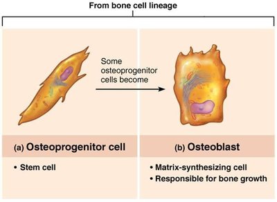
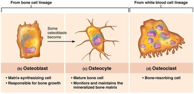
Osteogenic cells: Stem cells in periosteum and endosteum; differentiate into osteoblasts or bone-lining cells.
Osteoblasts: Bone-forming cells; secrete osteoid (collagen and calcium-binding proteins).
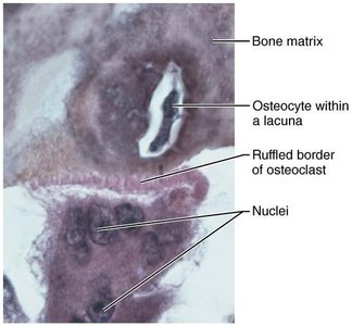
Osteocytes: Mature bone cells in lacunae; maintain matrix and act as stress sensors.
Bone-lining cells: Flat cells maintaining matrix; periosteal (external) and endosteal (internal).
Osteoclasts: Multinucleate cells for bone resorption; derived from hematopoietic stem cells.
Compact Bone
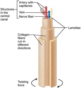
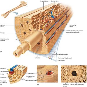
Osteon (Haversian system): Structural unit; elongated cylinder with concentric lamellae.
Lamellae: Collagen fibers run in different directions; resist twisting.
Canals and canaliculi: Central canal (blood vessels, nerves), perforating canals (connect periosteum, medullary cavity, central canal), canaliculi (connect lacunae).
Interstitial and circumferential lamellae: Fill gaps and extend around diaphysis.
Spongy Bone
Trabeculae align along lines of stress; confer strength.
No osteons; irregular lamellae and osteocytes interconnected by canaliculi.
Capillaries in endosteum supply nutrients.
Chemical Composition of Bone
Organic components: Bone cells and osteoid (ground substance and collagen fibers); provide tensile strength and flexibility.
Inorganic components: Hydroxyapatites (mineral salts, mainly calcium phosphate); provide hardness and resistance to compression.
Bone Development (Ossification)
Ossification is the process of bone tissue formation, beginning in the second month of development and continuing throughout life.
Formation of the Bony Skeleton
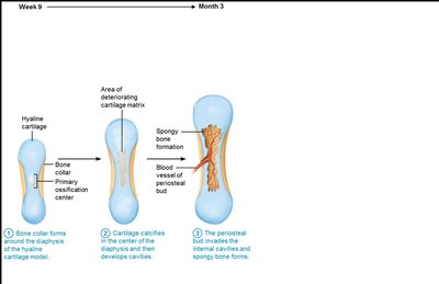
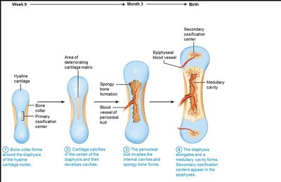
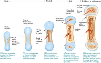
Endochondral ossification: Bone replaces hyaline cartilage; forms most bones below the skull.
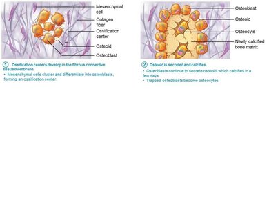
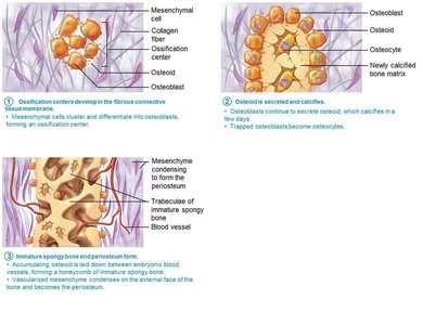
Intramembranous ossification: Bone develops from fibrous membrane; forms skull and clavicle bones.
Endochondral Ossification Steps
Bone collar forms around diaphysis of cartilage model.
Central cartilage in diaphysis calcifies, forming cavities.
Periosteal bud invades cavities, forming spongy bone.
Diaphysis elongates, medullary cavity forms; secondary ossification centers appear in epiphyses.
Epiphyses ossify; hyaline cartilage remains in epiphyseal plates and articular cartilages.
Intramembranous Ossification Steps
Ossification centers form as mesenchymal cells become osteoblasts.
Osteoid is secreted and calcified.
Woven bone forms around blood vessels, creating trabeculae; periosteum forms.
Lamellar bone replaces woven bone; red marrow appears.
Postnatal Bone Growth
Long bones grow in length by interstitial growth of the epiphyseal plate.
Bones increase in thickness by appositional growth.
Growth ceases during adolescence; some facial bones continue to grow slowly.
Epiphyseal Plate Zones
Resting zone: Inactive cartilage.
Proliferation zone: Rapidly dividing cartilage cells.
Hypertrophic zone: Older chondrocytes enlarge.
Calcification zone: Matrix calcifies, chondrocytes die.
Ossification zone: Calcified cartilage replaced by bone.
Hormonal Regulation of Bone Growth
Growth hormone: Stimulates epiphyseal plate activity.
Thyroid hormone: Modulates growth hormone activity.
Testosterone/Estrogen: Promote growth spurts and induce epiphyseal plate closure.
Bone Remodeling
Bone mass is recycled weekly; spongy bone every 3-4 years, compact bone every 10 years.
Remodeling involves bone deposit (by osteoblasts) and resorption (by osteoclasts).
Hormonal Controls
Parathyroid hormone (PTH): Stimulates osteoclasts to release calcium when blood levels are low.
Calcitonin: Lowers blood calcium levels at high doses.
Mechanical Stress
Wolff's law: Bones remodel in response to mechanical demands.
Bone thickens where stresses are greatest; trabeculae align along lines of stress.
Bone Repair
Fractures are classified by position, completeness, and skin penetration.
Repair involves four stages: hematoma formation, fibrocartilaginous callus formation, bony callus formation, and bone remodeling.
Bone Disorders
Osteomalacia and rickets: Poor mineralization; soft, weak bones due to vitamin D or calcium deficiency.
Osteoporosis: Bone resorption exceeds deposit; common in aged, postmenopausal women.
Paget’s disease: Excessive bone deposit and resorption; poorly formed bone.
Developmental Aspects and Age-Related Changes
Embryonic skeleton ossifies predictably; most long bones begin ossifying by 8 weeks.
Bone formation exceeds resorption in children; balanced in young adults; resorption exceeds formation in older adults.
Bone mass, mineralization, and healing decrease with age, especially in females and whites.
Additional info: These notes expand on brief points with academic context, definitions, and examples for self-contained study.
