 Indietro
Bones, Skeletal Anatomy, and Muscle Groups: A&P I Lab Practical 2 Study Guide
Guida di studio - Note intelligenti
 Appunti personalizzati basati sui tuoi materiali, ampliati con definizioni chiave, esempi e contesto.
Skull Anatomy
Cranium
The cranium consists of eight bones that protect the brain and provide attachment sites for head and neck muscles. Key features include foramina, sinuses, and processes that serve as passageways and attachment points.
Occipital bone: Contains the foramen magnum (passage for spinal cord), jugular foramen, and hypoglossal canal.
Parietal bones: Form the sides and roof of the cranium.
Frontal bone: Features the supraorbital foramen and frontal sinus.
Temporal bones: Include the zygomatic process, styloid process, mastoid process, and mandibular fossa.
Sphenoid bone: Contains the sella turcica, greater and lesser wings, and sphenoidal sinus.
Ethmoid bone: Features the cribriform plate, crista galli, and ethmoid air cells.
Face
The facial bones form the structure of the face, house the teeth, and provide passageways for nerves and blood vessels.
Maxillae: Contain the infraorbital foramen, alveolar processes, and maxillary sinus.
Zygomatic bones: Form the cheekbones and part of the zygomatic arch.
Mandible: Features the mental foramen, mandibular foramen, and condylar process.
Other bones: Palatine, nasal, lacrimal, inferior nasal conchae, and vomer.
Associated Bones
Hyoid bone: Supports the tongue and is not directly attached to other bones.
Auditory ossicles: Transmit sound vibrations in the middle ear.
Major Sutures of the Skull
Mid Sagittal Suture
Frontal/Coronal Suture
Lambdoid Suture
Squamous Suture
Vertebral Column
General Vertebrae Anatomy
The vertebral column consists of 26 vertebrae, each with distinct anatomical features. The vertebrae protect the spinal cord and provide structural support.
Vertebral body: Main weight-bearing region.
Vertebral foramen: Passage for the spinal cord.
Lamina and pedicle: Form the vertebral arch.
Spinous and transverse processes: Attachment points for muscles and ligaments.
Intervertebral foramen: Passage for spinal nerves (formed between adjacent vertebrae).
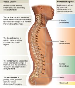
Vertebral Regions
Cervical (C1–C7): Includes the atlas (C1) and axis (C2), which allow head movement.
Thoracic (T1–T12): Articulate with ribs; have costal facets.
Lumbar (L1–L5): Largest vertebrae; support lower back.
Sacrum: Fused vertebrae forming the posterior pelvic wall.
Coccyx: Tailbone; vestigial structure.
Spinal Curves
The vertebral column has four curves that help maintain balance and absorb shock.
Cervical curve: Secondary curve; develops as infants lift their heads.
Thoracic curve: Primary curve; present at birth.
Lumbar curve: Secondary curve; develops as children begin to walk.
Sacral curve: Primary curve; present at birth.
Specialized Vertebrae
Atlas (C1): Supports the skull; allows nodding motion.
Axis (C2): Contains the dens; allows rotation of the head.
Thoracic Cage
Sternum and Ribs
The thoracic cage protects vital organs and supports the shoulder girdles and upper limbs.
Sternum: Composed of the manubrium, body, and xiphoid process.
Ribs: 12 pairs; classified as true (1–7), false (8–12), and floating (11–12).
Rib structure: Includes head, neck, tubercle, angle, and body.
Pectoral Girdle and Upper Limbs
Clavicle and Scapula
The pectoral girdle attaches the upper limbs to the trunk and provides mobility and stability.
Clavicle: S-shaped bone; articulates with sternum and scapula.
Scapula: Flat bone; features include acromion, coracoid process, glenoid cavity, and spine.
Upper Limb Bones
Humerus: Largest bone of the upper limb; articulates with scapula and forearm.
Radius and Ulna: Forearm bones; radius is lateral, ulna is medial.
Carpals, Metacarpals, Phalanges: Bones of the wrist, palm, and fingers.
Pelvic Girdle and Lower Limbs
Coxal Bone
The pelvic girdle supports the weight of the upper body and protects pelvic organs.
Ilium: Largest part; includes iliac crest and fossa.
Ischium: Features ischial tuberosity and ramus.
Pubis: Includes pubic symphysis and ramus.
Acetabulum: Socket for femur head.
Lower Limb Bones
Femur: Thigh bone; largest bone in the body.
Patella: Kneecap; protects knee joint.
Tibia and Fibula: Leg bones; tibia is medial and weight-bearing, fibula is lateral.
Tarsals, Metatarsals, Phalanges: Bones of the ankle, foot, and toes.
Muscle Groups and Movements
Major Muscle Groups
Muscle groups are classified by their location and function. Understanding their origins, insertions, and actions is essential for identifying movement and function.
Facial muscles
Mastication muscles
Rotator cuff muscles
Quadriceps and hamstrings
Gluteal group
Adductor group
Knee flexors and extensors
Ankle dorsiflexors and plantarflexors
Movements to Know
Flexion vs. Extension: Decreasing vs. increasing joint angle.
Abduction vs. Adduction: Moving away from vs. toward midline.
Lateral vs. Medial rotation: Rotating away from vs. toward midline.
Supination vs. Pronation: Rotating forearm to turn palm up vs. down.
Eversion vs. Inversion: Turning sole of foot outward vs. inward.
Dorsiflexion vs. Plantarflexion: Raising vs. lowering foot at ankle.
Retraction vs. Protraction: Moving a part backward vs. forward.
Depression vs. Elevation: Lowering vs. raising a body part.
Lateral flexion: Bending body sideways.
Skeletal Muscle Anatomy
Connective Tissue Layers
Skeletal muscles are organized into three connective tissue layers that provide structure and support.
Epimysium: Surrounds the entire muscle.
Perimysium: Surrounds muscle fascicles.
Endomysium: Surrounds individual muscle fibers.
Muscle Fiber Structure
Mitochondria: Provide energy for contraction.
Sarcoplasm: Cytoplasm of muscle fiber.
Sarcolemma: Plasma membrane of muscle fiber.
Myofibril: Contractile unit containing sarcomeres.
Sarcoplasmic reticulum: Stores calcium ions.
T-tubules: Transmit action potentials.
Nuclei: Multiple per fiber; control protein synthesis.
Sarcomere: Functional unit of contraction; composed of thin (actin) and thick (myosin) filaments.
Summary Table: Vertebral Regions and Curves
Region | Number of Vertebrae | Curve Type | Function |
|---|---|---|---|
Cervical | 7 | Secondary | Balance head, mobility |
Thoracic | 12 | Primary | Protect thoracic organs |
Lumbar | 5 | Secondary | Support lower back |
Sacral | 5 (fused) | Primary | Support pelvic organs |
Coccygeal | 4 (fused) | None | Vestigial |
