 Indietro
Cardiac Function and Regulation: Study Notes for Anatomy & Physiology
Guida di studio - Note intelligenti
 Appunti personalizzati basati sui tuoi materiali, ampliati con definizioni chiave, esempi e contesto.
Cardiac Function and the Cardiovascular System
Overview of Blood Flow Through the Cardiovascular System
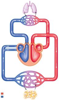
The cardiovascular system is responsible for the circulation of blood throughout the body, delivering oxygen and nutrients to tissues and removing waste products. Blood flows through two main circuits: the pulmonary circuit (to and from the lungs) and the systemic circuit (to and from the rest of the body).
Pulmonary Circuit: Carries deoxygenated blood from the right side of the heart to the lungs, where it becomes oxygenated.
Systemic Circuit: Distributes oxygenated blood from the left side of the heart to tissues throughout the body.
Oxygen Exchange: Occurs in capillary beds; oxygen moves into blood in pulmonary capillaries, and leaves blood in systemic capillaries.
Parallel and Series Blood Flow Patterns
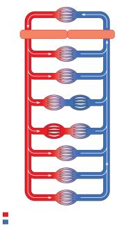
Blood flow in the systemic circuit is organized in parallel, allowing each organ to receive freshly oxygenated blood and enabling regulation of blood flow according to demand.
Parallel Flow: Most systemic tissues receive blood simultaneously, which helps regulate flow and prevents mixing of blood between organs.
Series Flow: Some exceptions exist, such as portal systems (e.g., hepatic portal system), where blood flows through two capillary beds in series.
Anatomy of the Heart
Structure of the Human Heart
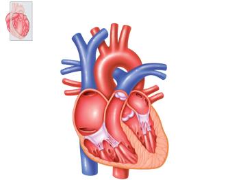
The heart is a muscular organ with four chambers: two atria and two ventricles. It contains valves that ensure unidirectional blood flow and prevent backflow.
Right Atrium and Ventricle: Receive deoxygenated blood and pump it to the lungs.
Left Atrium and Ventricle: Receive oxygenated blood and pump it to the systemic circuit.
Valves: Atrioventricular (AV) valves and semilunar valves control blood flow between chambers and out of the heart.
Electrical Activity of the Heart
Conduction System of the Heart
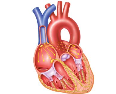
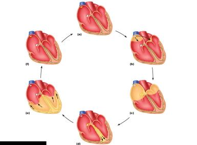
The heart's electrical activity is coordinated by specialized cells and pathways that generate and propagate action potentials, leading to rhythmic contractions.
Autorhythmic Cells: Generate spontaneous action potentials.
Pacemaker Cells: Located in the sinoatrial (SA) node, initiate the heartbeat.
Conduction Pathways: Include the SA node, atrioventricular (AV) node, bundle of His, bundle branches, and Purkinje fibers.
Gap Junctions: Allow electrical signals to spread rapidly between cells.
Spread of Action Potentials
Action potentials originate in the SA node and spread through the atria, AV node, and ventricles, ensuring coordinated contraction.
SA Node: Initiates action potential.
AV Node: Delays conduction to allow atrial contraction before ventricular contraction.
Bundle of His and Purkinje Fibers: Rapidly conduct action potentials to the ventricles.
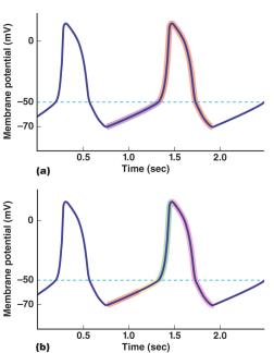
Action Potentials in Pacemaker Cells
Pacemaker cells exhibit unique action potentials characterized by slow depolarization (pacemaker potential), rapid depolarization, and repolarization.
Pacemaker Potential: Slow depolarization due to funny channels and T-type calcium channels.
Rapid Depolarization: L-type calcium channels open.
Repolarization: Potassium channels open.
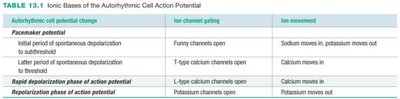
Autorhythmic cell potential change | Ion channel gating | Ion movement |
|---|---|---|
Initial period of spontaneous depolarization to subthreshold | Funny channels open | Sodium moves in, potassium moves out |
Later period of spontaneous depolarization to threshold | T-type calcium channels open | Calcium moves in |
Rapid depolarization phase of action potential | L-type calcium channels open | Calcium moves in |
Repolarization phase of action potential | Potassium channels open | Potassium moves out |
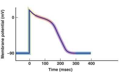
Action Potentials in Contractile Cells
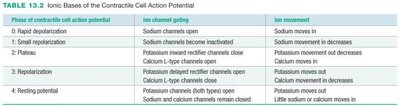
Contractile cells have action potentials with distinct phases: rapid depolarization, brief repolarization, plateau, and repolarization.
Depolarization: Sodium channels open.
Plateau: Calcium channels open, potassium channels close.
Repolarization: Potassium channels open, calcium channels close.
Phase of contractile cell action potential | Ion channel gating | Ion movement |
|---|---|---|
0: Rapid depolarization | Sodium channels open | Sodium moves in |
1: Small repolarization | Sodium channels become inactivated | Sodium movement in decreases |
2: Plateau | Potassium inward rectifier channels close, Calcium L-type channels open | Potassium movement out decreases, Calcium moves in |
3: Repolarization | Potassium delayed rectifier channels open, Calcium L-type channels close | Potassium moves out, Calcium movement in decreases |
4: Resting potential | Potassium channels both types open, Sodium and calcium channels remain closed | Little sodium or calcium moves in |
Mechanical Activity of the Heart
Cardiac Cycle
The cardiac cycle consists of alternating periods of contraction (systole) and relaxation (diastole) of the heart chambers, resulting in blood flow and pressure changes.
Systole: Ventricular contraction and ejection of blood.
Diastole: Ventricular relaxation and filling.
Heart Sounds: Produced by closure of heart valves.
Pressure-Volume Relationship in the Left Ventricle
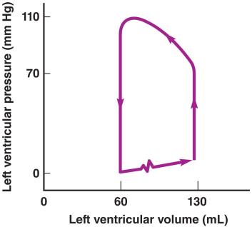
The pressure-volume curve illustrates the changes in ventricular pressure and volume during the cardiac cycle.
Isovolumetric Contraction: Pressure increases, volume remains constant.
Ventricular Ejection: Volume decreases as blood is pumped out.
Isovolumetric Relaxation: Pressure decreases, volume remains constant.
Ventricular Filling: Volume increases as blood enters the ventricle.
Key Cardiac Formulas
Mean Arterial Pressure (MAP):
Stroke Volume (SV):
Ejection Fraction (EF):
Cardiac Output (CO):
Regulation of Cardiac Function
Autonomic Regulation of Heart Rate and Force
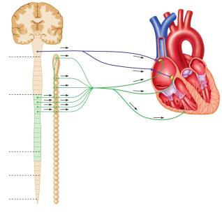
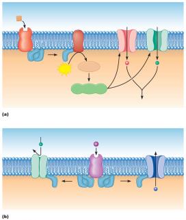
The autonomic nervous system regulates heart rate and contractile force through sympathetic and parasympathetic inputs.
Sympathetic Stimulation: Increases heart rate and force of contraction via norepinephrine and epinephrine.
Parasympathetic Stimulation: Decreases heart rate via acetylcholine.
Intrinsic Control: The heart can adjust its force of contraction based on venous return (Frank-Starling law).
Effects of Autonomic Nervous System on SA Node
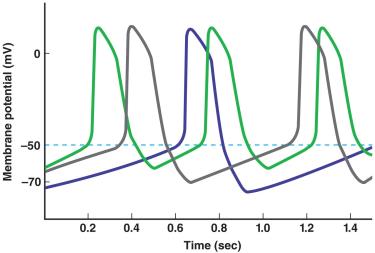
Sympathetic Activity: Increases rate of depolarization, raising heart rate.
Parasympathetic Activity: Increases potassium efflux, hyperpolarizing cells and slowing heart rate.
Sympathetic Regulation of Contractile Force
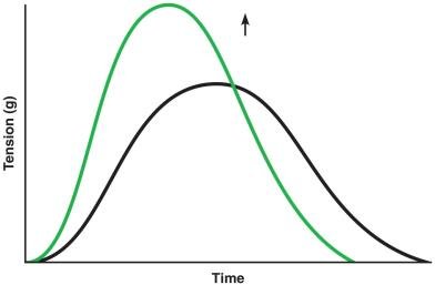
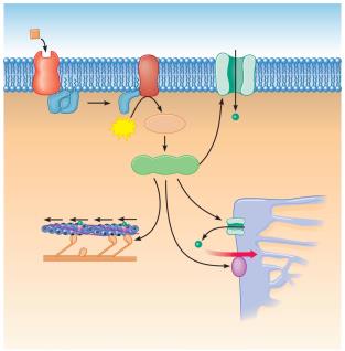
Sympathetic stimulation increases contractility by enhancing calcium influx and release, leading to stronger and faster contractions.
Norepinephrine/Epinephrine: Activate protein kinases, increase calcium channel opening.
Result: Increased crossbridge cycling and force generation.
Frank-Starling Law of the Heart
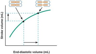
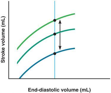
The Frank-Starling law states that an increase in end-diastolic volume (EDV) leads to an increase in stroke volume, allowing the heart to match output to venous return.
Optimal Sarcomere Length: Modest stretch increases force of contraction.
Independent of Neural/Hormonal Control: Intrinsic property of cardiac muscle.
Sympathetic Input and Frank-Starling Effect
Increased sympathetic activity shifts the Frank-Starling curve upward, increasing stroke volume at any given EDV.
Contractility: Enhanced by sympathetic stimulation.
Stroke Volume: Influenced by EDV, contractility, and afterload.
Summary Table: Factors Influencing Stroke Volume
Factor | Effect on Stroke Volume |
|---|---|
Venous Return | Increases EDV, increases SV |
Sympathetic Activity/Epinephrine | Increases contractility, increases SV |
Arterial Pressure (Afterload) | Increases afterload, decreases SV |
Key Terms and Concepts
Autorhythmic Cells: Specialized cardiac cells that generate action potentials without external stimulation.
Contractile Cells: Cardiac muscle cells responsible for force generation and pumping blood.
Pacemaker Potential: Slow depolarization in pacemaker cells leading to spontaneous action potentials.
Cardiac Cycle: Sequence of events in one heartbeat, including systole and diastole.
Stroke Volume (SV): Volume of blood ejected per beat.
Cardiac Output (CO): Total volume of blood pumped per minute.
Mean Arterial Pressure (MAP): Average pressure in arteries during one cardiac cycle.
Frank-Starling Law: Relationship between EDV and SV.
Example Calculations
MAP Calculation: If systolic BP = 140 mmHg, diastolic BP = 80 mmHg:
Stroke Volume: If EDV = 130 mL, ESV = 60 mL:
Ejection Fraction:
Cardiac Output: If HR = 75 bpm, SV = 70 mL:
Conclusion
Understanding cardiac function involves integrating knowledge of heart anatomy, electrical activity, mechanical function, and regulatory mechanisms. Mastery of these concepts is essential for comprehending cardiovascular physiology and its clinical applications.
