 Indietro
Cardiovascular Physiology: Electrical and Mechanical Events of the Heart
Guida di studio - Note intelligenti
 Appunti personalizzati basati sui tuoi materiali, ampliati con definizioni chiave, esempi e contesto.
Cardiac Anatomy and Blood Circulation
Internal Gross Anatomy of the Heart
The heart is a muscular organ responsible for pumping blood throughout the body. Its internal structure includes four chambers: two atria and two ventricles, as well as valves that regulate blood flow. - Atria: Upper chambers that receive blood. - Ventricles: Lower chambers that pump blood out. - Valves: Ensure unidirectional flow (AV valves and semilunar valves). 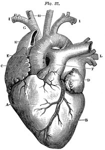
Blood Circulation Routes & Cardiac Cycle
Blood flows through two main circuits: pulmonary (to the lungs) and systemic (to the body). The cardiac cycle consists of alternating periods of contraction (systole) and relaxation (diastole). - Pulmonary circuit: Right side pumps blood to lungs for oxygenation. - Systemic circuit: Left side pumps oxygenated blood to the body.
Types of Cardiac Cells and Electrical Conduction
Types of Cardiac Cells
The heart contains two main types of cells: autorhythmic cells (pacemaker cells) and contractile cells. - Autorhythmic cells: Generate spontaneous action potentials. - Contractile cells: Respond to electrical signals and contract to pump blood.
Electrical Conduction in Myocardial Cells
Electrical impulses originate in the SA node and spread through the heart via gap junctions, causing coordinated contraction. - SA node: Primary pacemaker. - AV node: Delays impulse, allowing atrial contraction before ventricular contraction. - Bundle branches & Purkinje fibers: Distribute impulse to ventricles. 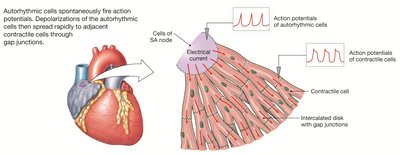
Myocardial Action Potentials
Action Potentials in Autorhythmic Cells
Autorhythmic cells have an unstable membrane potential called the pacemaker potential. - Depolarization: Initiated by Na+ inflow through If channels, followed by Ca2+ inflow. - Threshold: Second type of Ca2+ channels open, causing steep depolarization. - Repolarization: Ca2+ channels close, K+ channels open, returning to lowest potential. 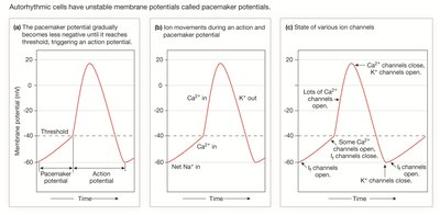
Action Potentials in Contractile Cells
Contractile cells have a distinct action potential with five phases:
Phase | Membrane Channels |
|---|---|
0 | Voltage-gated Na+ channels open |
1 | Voltage-gated Na+ channels close |
2 | Voltage-gated Ca2+ channels open; fast K+ channels close |
3 | Voltage-gated Ca2+ channels close; slow K+ channels open |
4 | Resting membrane potential |
Excitation-Contraction Coupling in Cardiac Muscle
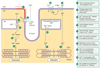
EC Coupling Mechanism
Excitation-contraction (EC) coupling links electrical signals to muscle contraction. - Action potential triggers Ca2+ entry. - Ca2+ release from sarcoplasmic reticulum (SR) amplifies signal. - Ca2+ binds to troponin, enabling contraction. - Relaxation occurs as Ca2+ is pumped back into SR and out of the cell. 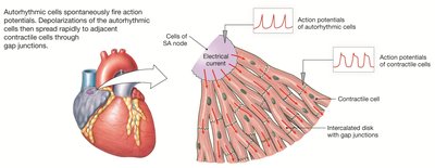
Refractory Periods in Cardiac Muscle
Cardiac Cell Refractory Period
The refractory period in cardiac muscle is as long as the action potential, preventing summation and tetanus. - Refractory period: Time during which a new action potential cannot be initiated. - Importance: Ensures rhythmic contraction and prevents sustained contraction (tetanus). 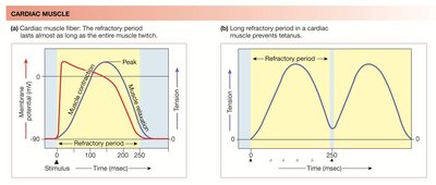
Conduction System of the Heart
Spread/Propagation of Contraction
The conduction system ensures coordinated contraction of the heart chambers. - SA node: Initiates depolarization. - AV node: Delays signal. - Bundle of His, bundle branches, Purkinje fibers: Rapidly distribute impulse to ventricles. 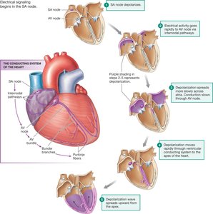
Electrocardiogram (ECG) and Cardiac Arrhythmias
ECG Waves and Segments
The ECG records the summed electrical activity of the heart. - P wave: Atrial depolarization. - QRS complex: Ventricular depolarization (includes atrial repolarization). - T wave: Ventricular repolarization. - P-R segment: AV nodal delay. - T-P segment: Ventricular and atrial relaxation. 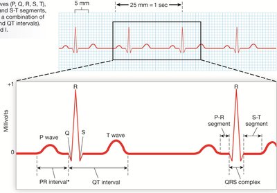
ECG Analysis and Arrhythmias
ECG analysis involves assessing heart rate, rhythm, wave presence, and QRS-P relationships. - Bradycardia: Heart rate < 60 bpm. - Tachycardia: Heart rate > 100 bpm. - Fibrillation: Asynchronous contraction, "bag of worms" appearance. 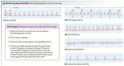
Cardiac Cycle: Mechanical Events
Phases of the Cardiac Cycle
The cardiac cycle consists of five phases: 1. Atrial and ventricular diastole: Heart at rest, AV valves open, ventricles fill. 2. Atrial systole: Atria contract, last 20% of blood driven to ventricles (EDV). 3. Early ventricular contraction: AV valves close, "Lub" sound, isovolumic contraction. 4. Ventricular ejection: Semilunar valves open, blood ejected, ESV measured. 5. Ventricular relaxation: Semilunar valves shut, "Dup" sound, isovolumic relaxation. 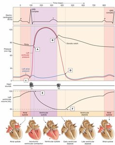
Stroke Volume and Cardiac Output
Stroke Volume
Stroke volume (SV) is the volume of blood pumped per contraction. - Formula: - Average: 70 mL (at rest)
Cardiac Output
Cardiac output (CO) is the volume of blood pumped by one ventricle per minute. - Formula: - Average: 5 L/min
Factors Influencing Stroke Volume
Contractility and Frank-Starling Law
- Contractility: Intrinsic ability of cardiac muscle to contract at any fiber length. - Frank-Starling law: Greater stretch (preload) before contraction results in stronger contraction. - Venous return: Influenced by skeletal muscle pump, respiratory pump, and sympathetic innervation. 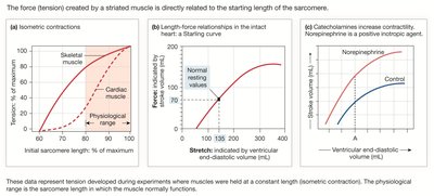
Regulation of Cardiac Function
Inotropic and Chronotropic Effects
- Inotropic agents: Chemicals affecting contractility (e.g., epinephrine, norepinephrine, digitalis). - Positive inotropes: Increase contractility. - Negative inotropes: Decrease contractility. - Chronotropic effects: Affect heart rate via autorhythmic cells. 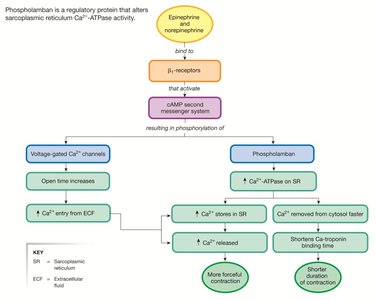
Parasympathetic and Sympathetic Innervation
- Parasympathetic NS: Slows heart rate via SA and AV node, no effect on contractility. - Sympathetic NS: Increases heart rate and contractility. 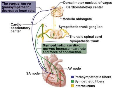
Summary Table: Cardiac Output Regulation
Factor | Effect on Cardiac Output |
|---|---|
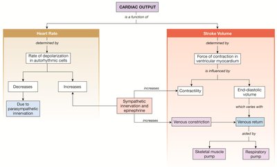
Heart Rate | Determined by rate of depolarization in autorhythmic cells; decreased by parasympathetic, increased by sympathetic innervation |
Stroke Volume | Determined by force of contraction in ventricular myocardium; influenced by contractility and end-diastolic volume |
Venous Return | Aided by skeletal muscle pump, respiratory pump, and sympathetic innervation |
Key Equations
Stroke Volume:
Cardiac Output:
Additional info:
Cardiac muscle's long refractory period is essential for preventing tetanus and ensuring rhythmic contractions.
Phospholamban is a regulatory protein that modulates Ca2+ handling in cardiac muscle, affecting contractility.
