 Indietro
Cardiovascular Physiology: Structure and Function of the Heart
Guida di studio - Note intelligenti
 Appunti personalizzati basati sui tuoi materiali, ampliati con definizioni chiave, esempi e contesto.
Cardiovascular System Overview
Components and Functions
The cardiovascular system is essential for transporting materials throughout the body. It consists of the heart (the pump), blood (the fluid), and blood vessels (the tubes). The system moves nutrients, water, gases, hormones, immune cells, and antibodies, and removes waste products such as carbon dioxide (CO2).
Heart: Central pump divided into left and right halves by the septum.
Blood Vessels: Includes arteries, veins, and capillaries. Arteries carry blood away from the heart; veins return blood to the heart.
Blood: Composed of cells and plasma.
Circulation: Pulmonary (to lungs) and systemic (to body tissues).
Internal Anatomy of the Heart
Chambers and Major Vessels
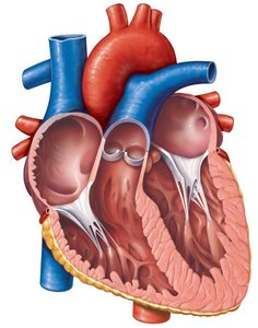
The heart has four chambers: right atrium, right ventricle, left atrium, and left ventricle. The septum divides the heart into left and right sides, preventing mixing of oxygenated and deoxygenated blood. Blood enters the atria and exits through the ventricles.
Atria: Receive blood returning to the heart (right: vena cavae; left: pulmonary veins).
Ventricles: Pump blood out of the heart (right: pulmonary trunk; left: aorta).
Septa: Keep blood from mixing between circuits.
Blood Circulation Routes
Pulmonary and Systemic Circuits
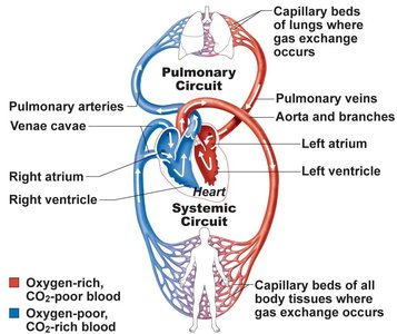
Blood flows through two parallel circuits: pulmonary and systemic. The right side of the heart receives deoxygenated blood and sends it to the lungs (pulmonary circuit), while the left side receives oxygenated blood from the lungs and sends it to the body (systemic circuit).
Pulmonary Circuit: Deoxygenated blood enters the right atrium via vena cavae, passes to the right ventricle, and is pumped to the lungs via the pulmonary trunk.
Systemic Circuit: Oxygenated blood returns to the left atrium via pulmonary veins, passes to the left ventricle, and is pumped to the body via the aorta.
The Heart as a Pump
Cardiac Cycle and Valve Function
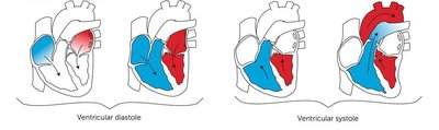
The cardiac cycle consists of alternating contraction (systole) and relaxation (diastole) of the atria and ventricles, ensuring unidirectional blood flow. Valves prevent backflow: atrioventricular (AV) valves between atria and ventricles, and semilunar (SL) valves between ventricles and major arteries.
Chordae Tendinae: "Heart strings" anchor AV valves, preventing inversion.
Valve Types: Tricuspid (right AV), bicuspid/mitral (left AV), aortic and pulmonary SL valves.
Anastomosis: Alternative routes for blood flow, especially in coronary circulation.
Cardiac Muscle and the Heart
Types of Cardiac Cells
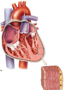
Cardiac muscle is composed of two main cell types: contractile cells (99%) and autorhythmic cells (1%). Contractile cells generate force for pumping blood, while autorhythmic cells initiate and propagate electrical impulses.
Contractile Cells: Striated, organized into sarcomeres, form the myocardium, and are connected via intercalated discs.
Autorhythmic Cells: Non-contractile, self-generate action potentials, located in the SA and AV nodes, and act as pacemakers.
Structural Features of Cardiac Muscle
Cardiac myocytes are striated and connected by intercalated discs, which contain desmosomes (mechanical connection) and gap junctions (electrical connection). Gap junctions allow rapid spread of action potentials, enabling synchronized contraction of heart chambers.
Desmosomes: Provide mechanical strength.
Gap Junctions: Allow electrical coupling for coordinated contraction.
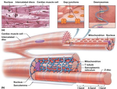
Comparison: Skeletal vs. Cardiac Muscle Cells
Feature | Skeletal Muscle | Cardiac Muscle |
|---|---|---|
Nuclei | Multinucleated | 1-2 nuclei |
Fiber Length | Long as entire muscle | Short, branched |
Contraction | Voluntary, somatic NS | Involuntary, ANS modifies rate |
AP Duration | 1-2 ms | 200-300 ms |
Coupling | NMJ | Intercalated discs |
Metabolism | Anaerobic/aerobic | Exclusively aerobic |
Conduction System of the Heart
Autorhythmic Cells and Electrical Pathways
Autorhythmic cells, also known as pacemaker cells, are responsible for initiating and propagating the electrical impulses that trigger heart contraction. The main components of the conduction system are the Sinoatrial (SA) Node, Atrioventricular (AV) Node, Bundle of His, and Purkinje fibers.
SA Node: Located in the right atrium; initiates the heartbeat.
AV Node: Electrical gateway between atria and ventricles.
Bundle of His and Purkinje Fibers: Distribute the impulse throughout the ventricles.
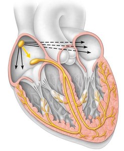
Spread and Propagation of Contraction
Electrical impulses generated by the SA node spread through the atria, reach the AV node, and then propagate through the bundle fibers and Purkinje fibers to the ventricles. This ensures coordinated contraction of the heart chambers.
Atrial and Ventricular Contraction: Atria contract first, followed by ventricles.
Gap Junctions: Enable rapid depolarization and synchronized contraction.
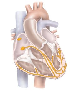
Summary Table: Cardiac Cell Types
Cell Type | Function | Location |
|---|---|---|
Contractile Cells | Generate force for pumping blood | Myocardium |
Autorhythmic Cells | Initiate and propagate action potentials | SA and AV nodes |
Key Equations
Cardiac Output
Cardiac output is the volume of blood pumped by the heart per minute.
Formula:
Stroke Volume
Stroke volume is the amount of blood ejected by a ventricle during each contraction.
Formula:
Conclusion
The cardiovascular system is a complex network responsible for the transport of essential materials and the removal of waste. The heart's structure, cell types, and conduction system ensure efficient and coordinated pumping of blood throughout the body.
