 Indietro
Cardiovascular System: Structure, Function, and Pathology
Guida di studio - Note intelligenti
 Appunti personalizzati basati sui tuoi materiali, ampliati con definizioni chiave, esempi e contesto.
Cardiovascular System Overview
Components and Functions
The cardiovascular system consists of the heart, blood vessels, and blood. It is responsible for transporting oxygen, nutrients, hormones, and waste products throughout the body, maintaining homeostasis, and supporting cellular function.
Heart: Pumps blood through the circulatory circuits.
Arteries: Carry blood away from the heart.
Veins: Carry blood toward the heart.
Blood: Connective tissue composed of plasma and formed elements (erythrocytes, leukocytes, platelets).
Circulatory Circuits
Pulmonary and Systemic Circuits
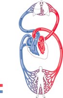
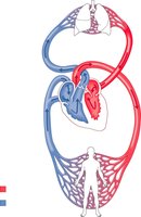
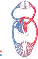
The heart pumps blood through two main circuits: the pulmonary circuit and the systemic circuit. These circuits ensure oxygenation of blood and distribution to body tissues.
Pulmonary Circuit: Carries blood from the right ventricle to the lungs for oxygenation and returns it to the left atrium.
Systemic Circuit: Distributes oxygen-rich blood from the left ventricle to the body and returns oxygen-poor blood to the right atrium.
Heart Structure and Location
Basic Anatomy and Chambers
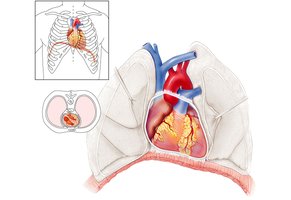
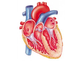
The heart is a muscular organ located slightly left of the midline, above the diaphragm, and enclosed in the pericardium. It has four chambers: right atrium, right ventricle, left atrium, and left ventricle.
Right Atrium: Receives deoxygenated blood from the body.
Right Ventricle: Pumps blood to the lungs.
Left Atrium: Receives oxygenated blood from the lungs.
Left Ventricle: Pumps oxygenated blood to the body.
Pericardium and Heart Wall Layers
The heart is surrounded by the pericardium, which consists of fibrous and serous layers. The heart wall itself has three layers: epicardium, myocardium, and endocardium.
Fibrous Pericardium: Tough outer layer anchoring the heart.
Serous Pericardium: Parietal (lines sac) and visceral (covers heart, also called epicardium) layers.
Myocardium: Cardiac muscle tissue responsible for contraction.
Endocardium: Simple squamous epithelium lining the chambers.
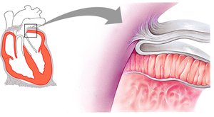
Heart Valves and Blood Flow
Atrioventricular and Semilunar Valves
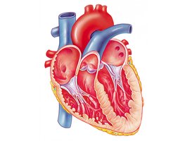
Valves ensure unidirectional blood flow through the heart. The atrioventricular (AV) valves separate atria from ventricles, while semilunar valves separate ventricles from arteries.
Tricuspid Valve: Right AV valve, separates right atrium and ventricle.
Bicuspid (Mitral) Valve: Left AV valve, separates left atrium and ventricle.
Pulmonary Semilunar Valve: Separates right ventricle and pulmonary trunk.
Aortic Semilunar Valve: Separates left ventricle and aorta.
Valve Function and Pathology
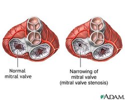
Valves open and close in response to pressure changes. Chordae tendineae and papillary muscles prevent valve prolapse. Valve defects can cause murmurs or stenosis.
Mitral Valve Prolapse: Weakness in valve or chordae tendineae causes blood leakage.
Stenosis: Narrowed valve opening, often due to calcium deposits or illness.
Cardiac Muscle and Conducting System
Microscopic Anatomy
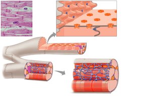
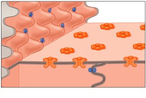
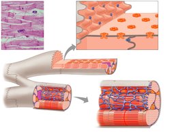
Cardiac muscle cells are striated and connected by intercalated discs, which contain gap junctions and fascia adherens for coordinated contraction and strength.
Intercalated Discs: Specialized junctions for electrical and mechanical connectivity.
Gap Junctions: Allow rapid communication between cells.
Fascia Adherens: Desmosome-like connections for structural integrity.
Conducting System of the Heart
The heart's conducting system initiates and propagates electrical impulses for rhythmic contraction. Key components include the sinoatrial (SA) node, atrioventricular (AV) node, AV bundle, bundle branches, and Purkinje fibers.
SA Node: Pacemaker, initiates impulse in right atrium.
AV Node: Delays impulse before passing to ventricles.
AV Bundle (Bundle of His): Only pathway from atria to ventricles.
Bundle Branches and Purkinje Fibers: Distribute impulse to ventricular muscle.
Blood Vessels: Structure and Function
Layers of Vessel Walls
Blood vessels are composed of three main layers: tunica intima, tunica media, and tunica externa. These layers vary in thickness and composition between arteries, veins, and capillaries.
Tunica Intima: Endothelium (simple squamous epithelium) and subendothelial layer (loose areolar CT).
Tunica Media: Smooth muscle, allows for vasoconstriction and vasodilation.
Tunica Externa: Loose areolar CT, contains vaso vasorum (vessels supplying vessel wall).
Arteries, Veins, and Capillaries
Arteries have thicker walls and more elastic fibers than veins, which have valves to prevent backflow. Capillaries are thin-walled for efficient exchange of gases and nutrients.
Arteries: High pressure, thick tunica media, elastic fibers.
Veins: Lower pressure, valves, thinner walls.
Capillaries: Single layer of endothelium, three types (continuous, fenestrated, sinusoidal).
Blood: Composition and Function
Components of Blood
Blood is a connective tissue composed of plasma and formed elements. It transports gases, nutrients, wastes, hormones, and regulates temperature and pH.
Plasma: Extracellular matrix, ~55% of blood volume, contains water, proteins (albumin, fibrinogen), solutes.
Formed Elements: Erythrocytes (RBCs), leukocytes (WBCs), platelets.
Erythrocytes (Red Blood Cells)
Erythrocytes are anucleate, biconcave cells specialized for oxygen transport via hemoglobin. They have a lifespan of 3-4 months and are produced in red bone marrow.
Hemoglobin: Protein that binds oxygen and carbon dioxide.
Hematopoiesis: Formation of new RBCs from stem cells; iron is essential for hemoglobin synthesis.
Leukocytes (White Blood Cells) and Platelets
Leukocytes are complete cells involved in immune response, capable of diapedesis. Platelets are cell fragments essential for blood clotting.
Leukocytes: Fight infection, increase in number during immune response.
Platelets: Aggregate to form clots and prevent bleeding.
Blood Groups and Rh Factor
ABO Blood Groups
Blood groups are determined by antigens (agglutinogens) on erythrocyte membranes. The main groups are A, B, AB, and O, each with specific antibodies produced against foreign antigens.
Type A: A antigens, produces anti-B antibodies.
Type B: B antigens, produces anti-A antibodies.
Type AB: Both A and B antigens, no antibodies (universal receiver).
Type O: No A or B antigens, produces both anti-A and anti-B antibodies (universal donor).
Rh Factor
The Rh factor is a protein that may be present (+) or absent (-) on erythrocytes. Rh antibodies can cross the placenta and affect fetal health, especially during birth if the mother is Rh-negative and the fetus is Rh-positive.
Rh-positive: Has Rh antigen.
Rh-negative: Lacks Rh antigen; can produce anti-Rh antibodies upon exposure.
Clinical Correlations
Heart Defects and Pathologies
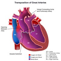
Congenital heart defects, such as transposition of the great arteries, can severely impact blood flow and oxygenation. Atherosclerosis and myocardial infarction are common cardiovascular diseases.
Transposition of Great Arteries: Aorta and pulmonary trunk are switched, leading to improper oxygenation.
Atherosclerosis: Plaque buildup narrows arteries, leading to reduced blood flow.
Myocardial Infarction: Heart attack due to blocked blood supply to cardiac muscle.
Anemia
Anemia is a condition characterized by a lower than normal number of red blood cells, resulting in reduced oxygen transport. It can be caused by iron deficiency, chronic disease, or nutritional deficits.
Iron Deficiency: Leads to fewer RBCs and less hemoglobin.
Symptoms: Fatigue, weakness, pallor.
Varicose Veins
Varicose veins occur when venous valves fail, causing blood to pool and veins to become misshapen. Most commonly affects superficial veins of the lower leg.
Valve Failure: Blood pools, stretches veins.
Symptoms: Swelling, pain, visible twisted veins.
Summary Table: Blood Vessel Structure
Vessel Type | Tunica Intima | Tunica Media | Tunica Externa | Special Features |
|---|---|---|---|---|
Artery | Endothelium, subendothelial layer | Thick, smooth muscle, elastic fibers | Moderate thickness, vaso vasorum | High pressure, rounded lumen |
Vein | Endothelium, valves | Thin, less smooth muscle | Thick, vaso vasorum | Low pressure, irregular lumen |
Capillary | Endothelium only | None | None | Exchange of gases/nutrients |
Additional info: Academic context was added to clarify the function and clinical relevance of cardiovascular structures and pathologies.
