 Indietro
Cell Membrane Structure, Transport Mechanisms, and Neuron Anatomy: Study Notes for Anatomy & Physiology
Guida di studio - Note intelligenti
 Appunti personalizzati basati sui tuoi materiali, ampliati con definizioni chiave, esempi e contesto.
Cell Membrane Structure and Function
Phospholipid Bilayer: The Fluid Mosaic Model
The plasma membrane is primarily composed of a phospholipid bilayer, which forms the basic structural framework of all cell membranes.
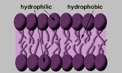
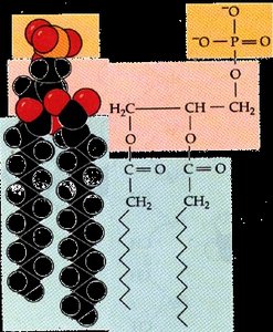
Phospholipids are amphipathic molecules, meaning they have both hydrophilic (water-loving) heads and hydrophobic (water-fearing) tails.
The hydrophilic heads face outward toward the aqueous environments inside and outside the cell, while the hydrophobic tails face inward, away from water.
This arrangement creates a selectively permeable barrier, allowing the cell to regulate its internal environment.
Components of the Plasma Membrane
The plasma membrane contains more than just phospholipids. Several other components contribute to its structure and function:
Integral membrane proteins: Span the membrane and have hydrophilic and hydrophobic regions. They function as channels, carriers, and receptors.
Peripheral proteins: Attached to integral proteins, often on the internal face of the membrane. They can act as enzymes, participate in attachment functions, or serve as receptors.
Cytoskeleton: Anchors to the plasma membrane, providing structural support.
Glycocalyx: A mix of carbohydrates attached to lipids and proteins on the outer face, forming a "sugar coating" that allows cell recognition.
Cholesterol: Stabilizes membrane structure and reduces fluidity; excessive cholesterol can decrease membrane flexibility.
Cell Junctions
Cell junctions are specialized structures that connect adjacent cells and serve various functions:
Tight junctions: Fusion of adjacent membranes to prevent passage of molecules between cells. Important in tissues like the digestive tract.
Desmosomes: Anchoring junctions that link cells to resist mechanical stress, found in tissues subject to stretching.
Gap junctions: Channels that allow passage of cytoplasmic molecules between cells, crucial in electrically excitable tissues.
Membrane Transport Mechanisms
Passive Transport
Passive transport does not require energy and relies on the movement of molecules down their concentration gradients.
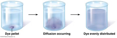
Diffusion: The tendency of molecules to scatter evenly throughout the environment due to kinetic energy. Rate is influenced by concentration gradient, molecule size, and temperature.
Simple diffusion: Lipid-soluble, nonpolar molecules (e.g., O2, CO2, fats, alcohol) pass directly through the membrane.
Facilitated diffusion: Water-soluble substances require carrier or channel proteins to cross the membrane. This process is specific, not ATP-requiring, and limited by carrier/channel saturation.
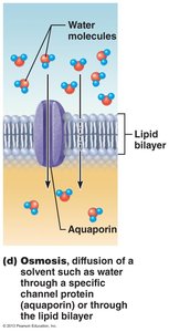
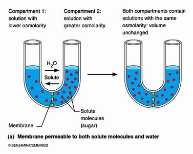
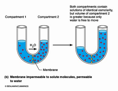
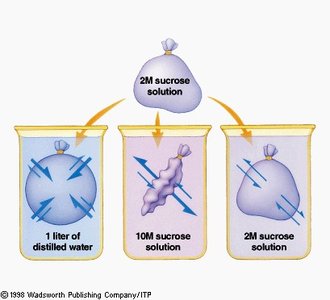
Osmosis: The unassisted diffusion of water across a semipermeable membrane from an area of higher water concentration to lower water concentration. Water moves through aquaporins or directly through the lipid bilayer.
Osmolarity and Tonicity
Osmolarity: Total concentration of solute particles in a solution, measured in mOsmol/L.
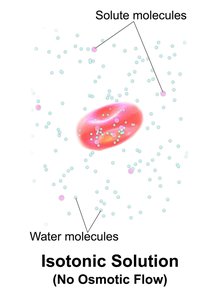
Tonicity: The ability of a solution to change the shape of a cell by altering its water content, determined by nonpenetrating solute particles.
Solutions are described as isotonic (no net water movement), hypertonic (water moves out of the cell), or hypotonic (water moves into the cell).
Active Transport
Active transport requires ATP to move substances against their concentration gradients or when substances are too large or lipid-insoluble.
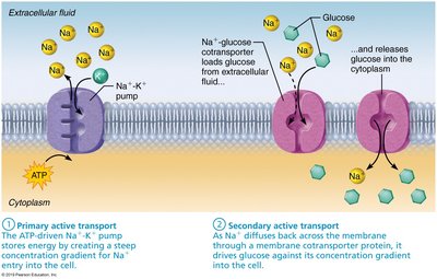
Primary active transport: Direct use of ATP, e.g., Na+/K+ ATPase pump, which maintains essential ion gradients by pumping 3 Na+ out and 2 K+ in.
Secondary active transport: Uses the gradient established by primary active transport to drive the movement of other substances (e.g., cotransport of glucose with Na+).
Transport systems can be symport (same direction) or antiport (opposite directions).
Vesicular Transport
Vesicular transport involves the movement of large particles or volumes of fluid via vesicles and requires ATP.
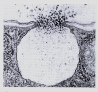
Exocytosis: The process by which cells secrete substances (e.g., hormones, neurotransmitters) by enclosing them in vesicles that fuse with the plasma membrane and release contents outside the cell.
Endocytosis: The process by which cells take in large particles or fluids. Includes phagocytosis (for solids), pinocytosis (for fluids), and receptor-mediated endocytosis (for specific macromolecules).
Neuron Structure and Function
Overview of Neurons
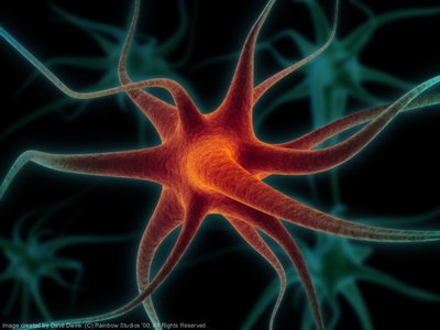
Neurons are specialized cells that conduct electrical impulses throughout the body.
They exhibit extreme longevity, are generally amitotic (do not divide), and have high metabolic rates requiring constant supply of O2 and glucose.
Each neuron consists of a cell body and one or more processes (dendrites and axons).

Functional Regions of the Neuron
Receptive region: Includes dendrites and cell body; receives signals from other neurons.
Conducting region: The axon; propagates electrical impulses away from the cell body.
Secretory region: Axon terminals; release neurotransmitters to communicate with other cells.
Neuron Cell Body and Processes
Cell body: Contains the nucleus and organelles; biosynthetic center of the neuron.
Dendrites: Short, branched extensions; receive signals and conduct impulses toward the cell body.
Axon: Long process; conducts impulses away from the cell body. Axon terminals release neurotransmitters.
Bundles of axons are called tracts in the CNS and nerves in the PNS.
Axonal Transport
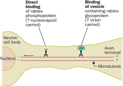
Materials are moved along the axon via the cytoskeleton in two directions: anterograde (toward axon terminals) and retrograde (toward cell body).
Viruses and toxins can exploit retrograde transport to reach the neuron cell body.
Summary Table: Types of Membrane Transport
Transport Type | Energy Required | Direction | Example |
|---|---|---|---|
Simple Diffusion | No | Down gradient | O2, CO2 |
Facilitated Diffusion | No | Down gradient | Glucose, ions |
Osmosis | No | Down gradient | Water |
Primary Active Transport | Yes (ATP) | Against gradient | Na+/K+ pump |
Secondary Active Transport | Indirect (ATP) | Against gradient | Na+-glucose cotransport |
Vesicular Transport | Yes (ATP) | Bulk movement | Exocytosis, endocytosis |
Key Equations
Osmolarity Calculation
Osmolarity is calculated as the sum of all solute particles in solution:
Example: 1 mM NaCl = 2 mOsm (Na+ and Cl-), 1 mM CaCl2 = 3 mOsm (Ca2+ and 2 Cl-).
Na+/K+ ATPase Pump
For every cycle:
This maintains the electrochemical gradient essential for cell function.
Clinical Applications
Hypertonic solutions can be used to treat edema by pulling water out of tissues.
Hypotonic solutions are used to rehydrate severely dehydrated patients.
Understanding membrane transport is crucial for interpreting physiological and pathological states.
Additional info:
Expanded explanations of membrane structure and transport mechanisms were added for completeness.
Clinical notes and examples were inferred from standard Anatomy & Physiology context.
