 Indietro
Central Nervous System: Structure and Function of the Brain and Spinal Cord
Guida di studio - Note intelligenti
 Appunti personalizzati basati sui tuoi materiali, ampliati con definizioni chiave, esempi e contesto.
Central Nervous System Overview
Introduction
The central nervous system (CNS) is composed of the brain and spinal cord. It is responsible for integrating sensory information, coordinating bodily functions, and facilitating higher mental processes such as thought, memory, and emotion. Understanding the structure and function of the CNS is essential for comprehending neurological health and disease.
Major Regions of the Brain
Brain Structure and Regions
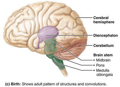
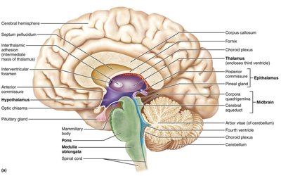
The adult brain is divided into four major regions: the cerebral hemispheres, diencephalon, brain stem, and cerebellum. Each region has distinct anatomical features and specialized functions.
Cerebral Hemispheres: The largest part of the brain, responsible for higher cognitive functions, voluntary movement, and sensory perception.
Diencephalon: Contains structures such as the thalamus and hypothalamus, which relay sensory information and regulate autonomic functions.
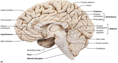
Brain Stem: Composed of the midbrain, pons, and medulla oblongata, it controls vital autonomic functions and connects the brain to the spinal cord.
Cerebellum: Coordinates movement and balance.
Gray Matter and White Matter
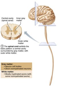
Gray matter consists of neuron cell bodies and nonmyelinated fibers, while white matter is composed mainly of myelinated axons. The basic pattern in the CNS is a central cavity surrounded by gray matter, with white matter external to it. This pattern is evident in the spinal cord and modified in the brain.
Ventricles of the Brain
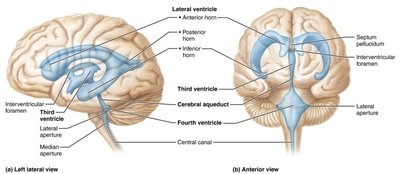
The brain contains interconnected, fluid-filled cavities called ventricles. These are lined by ependymal cells and filled with cerebrospinal fluid (CSF), which cushions and nourishes the brain.
Lateral Ventricles: Paired, C-shaped chambers in each hemisphere.
Third Ventricle: Located in the diencephalon, connected to the lateral ventricles via the interventricular foramen.
Fourth Ventricle: Located between the brain stem and cerebellum, connected to the third ventricle by the cerebral aqueduct.
Surface Features of the Cerebrum
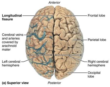
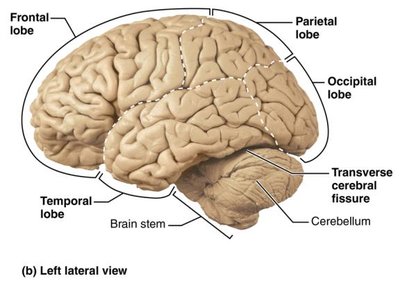
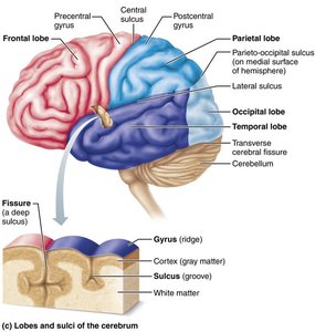
The surface of the cerebral hemispheres is marked by ridges (gyri), shallow grooves (sulci), and deep grooves (fissures). The longitudinal fissure separates the two hemispheres, while the transverse cerebral fissure separates the cerebrum from the cerebellum.
Lobes of the Cerebrum
Each hemisphere is divided into five lobes: frontal, parietal, temporal, occipital, and insula. The first four are named after the overlying skull bones, while the insula is buried beneath portions of the other lobes.
Functional Areas of the Cerebral Cortex
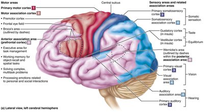
The cerebral cortex is the site of conscious mind activities, including awareness, sensory perception, voluntary movement, communication, memory, and understanding. It contains three main types of functional areas:
Motor Areas: Control voluntary movement.
Sensory Areas: Responsible for conscious awareness of sensation.
Association Areas: Integrate diverse information for complex functions.
Somatotopic Maps
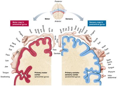
The primary motor cortex and primary somatosensory cortex are organized as somatotopic maps, representing different body regions. The size of each region on the map reflects the degree of motor control or sensory sensitivity.
Diencephalon
Thalamus, Hypothalamus, and Epithalamus
The diencephalon consists of three paired gray matter structures:
Thalamus: Relay station for sensory and motor signals to the cerebral cortex.
Hypothalamus: Main visceral control center, regulating homeostasis, autonomic functions, emotions, and the endocrine system.
Epithalamus: Contains the pineal gland, which secretes melatonin to regulate sleep-wake cycles.
Brain Stem
Structure and Function
The brain stem consists of the midbrain, pons, and medulla oblongata. It controls automatic behaviors necessary for survival and serves as a conduit for information between the brain and spinal cord.
Midbrain: Contains visual and auditory reflex centers.
Pons: Relays information between the cerebrum and cerebellum; involved in breathing regulation.
Medulla Oblongata: Regulates cardiovascular and respiratory functions, as well as reflexes such as swallowing and vomiting.
Cerebellum
Structure and Function
The cerebellum is located dorsal to the pons and medulla and accounts for about 11% of brain mass. It processes input from the cortex, brain stem, and sensory receptors to coordinate precise movements and maintain balance.
Spinal Cord
Structure of the Spinal Cord
The spinal cord is enclosed within the vertebral column, beginning at the foramen magnum and ending at the level of the first or second lumbar vertebra. It provides two-way communication between the brain and body and serves as a major reflex center.
Central Canal: Runs the length of the cord and is filled with CSF.
Gray Matter: Contains neuron cell bodies and is organized into dorsal, ventral, and lateral horns.
White Matter: Composed of ascending (sensory), descending (motor), and transverse (commissural) tracts.
Meninges and Cerebrospinal Fluid
Meninges
The meninges are three connective tissue membranes that cover and protect the CNS, contain cerebrospinal fluid, and form partitions within the skull. The layers are:
Dura Mater: The tough, outermost layer.
Arachnoid Mater: The middle, web-like layer.
Pia Mater: The delicate, innermost layer that adheres to the brain and spinal cord.
Cerebrospinal Fluid (CSF)
CSF is a clear, colorless fluid that cushions the brain and spinal cord, provides buoyancy, removes waste, and transports nutrients. It is produced by the choroid plexus in the ventricles and circulates through the ventricular system and subarachnoid space before being reabsorbed into the venous blood.
Clinical Implications
Stroke (Cerebrovascular Accident): Occurs when blood flow to a part of the brain is blocked, leading to tissue death and possible loss of function.
Hydrocephalus: Accumulation of CSF due to obstruction, causing increased intracranial pressure.
Meningitis: Inflammation of the meninges, which can spread to the CNS and cause encephalitis.
Spinal Cord Injury: Can result in loss of sensory and motor function below the level of injury (paraplegia or quadriplegia).
