 Indietro
Central Nervous System: Structure and Function
Guida di studio - Note intelligenti
 Appunti personalizzati basati sui tuoi materiali, ampliati con definizioni chiave, esempi e contesto.
Central Nervous System (CNS) Overview
Functions of the CNS
The central nervous system (CNS) consists of the brain and spinal cord and is responsible for integrating sensory information, coordinating motor output, and performing higher mental functions. It is the primary site for decision-making and regulation of bodily functions.
Motor functions: Initiate muscle contraction and gland secretion (primarily via the peripheral nervous system).
Sensory functions: Detect and interpret sensations from internal and external environments.
Integrative functions: Process and integrate information for complex decision-making (exclusive to the CNS).
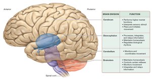
Major CNS Structures
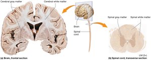
White Matter and Gray Matter
Both the brain and spinal cord contain white and gray matter, which differ in structure and function:
White matter: Composed of myelinated axons; responsible for transmitting signals rapidly across regions.
Gray matter: Contains neuron cell bodies, dendrites, and unmyelinated axons; involved in processing and integration of information.
The Cerebrum
Gross Anatomy of the Cerebrum
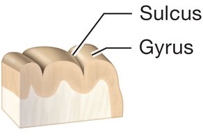
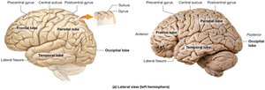
The cerebrum is the largest part of the brain, responsible for higher mental functions such as reasoning, memory, and voluntary movement. Its surface is characterized by distinct features:
Sulci: Shallow grooves on the surface.
Gyri: Elevated ridges between sulci.
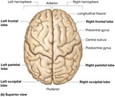
Fissures: Deep grooves, including the longitudinal fissure that separates the left and right hemispheres.
Lobes of the Cerebrum
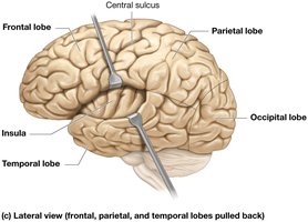
The cerebrum is divided into several lobes, each with specialized functions:
Frontal lobe: Motor function, problem-solving, speech production.
Parietal lobe: Sensory perception, spatial orientation.
Temporal lobe: Auditory processing, memory, language comprehension.
Occipital lobe: Visual processing.
Insula: Deep within the lateral sulcus; involved in taste, visceral sensation, and emotion.
Cerebral Cortex: Gray Matter
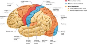
The cerebral cortex is the outer layer of gray matter and is functionally the most complex part of the brain. It is divided into three main areas:
Primary motor cortex: Initiates voluntary movements.
Primary sensory cortices: Receive and process sensory input.
Association areas: Integrate information; subdivided into unimodal (single function) and multimodal (multiple functions) areas.
Motor and Sensory Cortices
Specific regions of the cortex are dedicated to motor and sensory functions:
Premotor cortex: Plans movements.
Frontal eye fields: Control voluntary eye movements.
Somatosensory cortex: Processes touch, pain, and temperature.
Special senses cortices: Visual, auditory, gustatory, vestibular, and olfactory areas.
Multimodal Association Areas
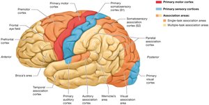
Multimodal association areas perform complex mental functions such as language, reasoning, and memory. Key regions include:
Broca’s area: Speech production.
Wernicke’s area: Language comprehension.
Prefrontal cortex: Executive functions, personality.
Parietal and temporal association areas: Integration of sensory information.
Cerebral White Matter
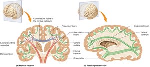
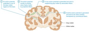
Cerebral white matter consists of three types of fibers:
Commissural fibers: Connect the two hemispheres (e.g., corpus callosum).
Projection fibers: Connect cortex with lower brain regions and spinal cord.
Association fibers: Connect regions within the same hemisphere.
Diencephalon
Major Components
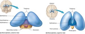
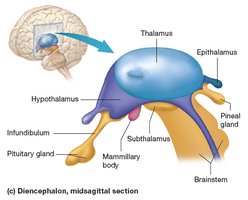
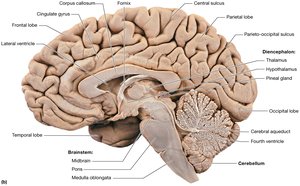
The diencephalon is located deep within the brain and consists of several key structures:
Thalamus: Relay station for sensory information.
Hypothalamus: Regulates homeostasis, links nervous and endocrine systems.
Mamillary bodies: Involved in memory.
Pituitary gland: Master endocrine gland.
Epithalamus: Contains pineal gland, regulates circadian rhythms.
Subthalamus: Motor control.
Cerebellum
Anatomy and Function
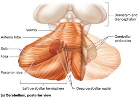
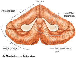
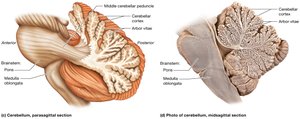
The cerebellum is responsible for monitoring and coordinating movement. It consists of two hemispheres connected by the vermis and is divided into three lobes: anterior, posterior, and flocculonodular. The cerebellar cortex is the outer layer of gray matter, and the arbor vitae is the internal white matter.
Folia: Folds on the cerebellar surface.
Cerebellar peduncles: Connect cerebellum to brainstem.
Brainstem
Structure and Function
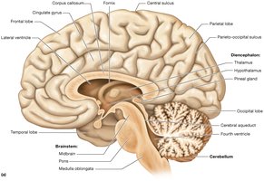
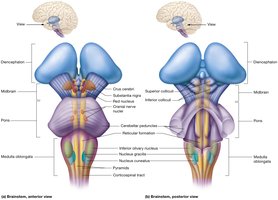
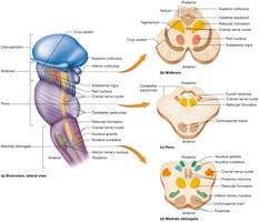
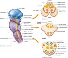
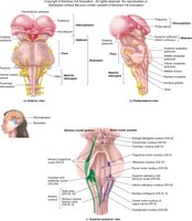
The brainstem is vital for survival, controlling basic homeostatic functions, reflexes, movement, sensation, and alertness. It is divided into three regions:
Midbrain: Contains corpora quadrigemina, superior and inferior colliculi, substantia nigra, red nucleus, and cranial nerve nuclei.
Pons: Contains descending motor tracts, pontine nuclei, and respiratory centers.
Medulla oblongata: Contains pyramids, olives, cardiovascular and respiratory centers, and cranial nerve nuclei.
Reticular Formation
Input and Output
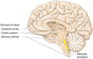
The reticular formation receives input from the cerebral cortex, limbic system, and sensory stimuli, and sends output throughout the brain and spinal cord. It is involved in sleep, pain transmission, mood, motor functions, and homeostasis.
Central nuclei: Sleep, pain, mood.
Surrounding nuclei: Motor functions.
Lateral nuclei: Sensation, alertness.
Brain Protection
Cranial Meninges and Structural Arrangement
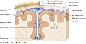
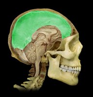
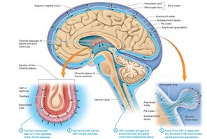
The brain is protected by three layers of meninges:
Dura mater: Tough, outermost layer (periosteal and meningeal layers).
Arachnoid mater: Middle layer with web-like trabeculae.
Pia mater: Delicate, innermost layer adhering to brain surface.
Spaces between these layers include the epidural, subdural, and subarachnoid spaces.
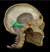
Inward Extensions of the Dura
Three major folds of dura mater provide additional protection:
Falx cerebri: Separates cerebral hemispheres.
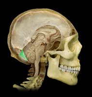
Tentorium cerebelli: Separates cerebrum from cerebellum.
Falx cerebelli: Separates cerebellar hemispheres.
The Ventricles and Cerebrospinal Fluid (CSF)
Ventricular System
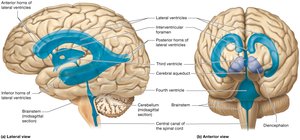
The brain contains four ventricles filled with cerebrospinal fluid (CSF):
Lateral ventricles: Located in each cerebral hemisphere.
Third ventricle: Between lobes of diencephalon.
Fourth ventricle: Between pons and cerebellum.
CSF Circulation
CSF is produced by the choroid plexus, circulates through ventricles, and is absorbed into the venous system. It cushions the brain and removes waste.
Blood Supply and Blood-Brain Barrier
Blood Supply to the Brain
The brain receives a significant portion of the body's blood supply due to its high demand for oxygen and glucose. Interruption of blood flow can quickly lead to loss of function and irreversible damage.
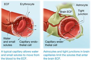
Blood-Brain Barrier
The blood-brain barrier protects the brain from harmful substances in the blood. It consists of endothelial cells, basal laminae, and astrocytes, allowing only certain substances to pass.
Small, nonpolar molecules: Pass easily.
Large, polar molecules: Are restricted.
The Spinal Cord
External Anatomy
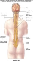
The spinal cord extends from the brain through the vertebral canal and is divided into cervical, thoracic, lumbar, and sacral regions. It gives rise to 31 pairs of spinal nerves.
Cervical and lumbosacral enlargements: Areas with increased nerve supply for limbs.
Conus medullaris: Tapered end of the spinal cord.
Cauda equina: Bundle of spinal nerves below the conus medullaris.
Filum terminale: Provides support to the spinal cord.
Protection of the Spinal Cord
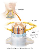
The spinal cord is protected by meninges similar to those in the brain, but with some structural differences:
Dura mater: Single layer in the spinal cord.
Pia mater: Has structural enhancements.
Spaces: Epidural, subdural, and subarachnoid spaces.
Internal Anatomy
Spinal gray matter is butterfly-shaped and surrounded by white matter. Key features include:
Central canal: Contains CSF.
Gray commissure: Connects the two sides of gray matter.
Anterior (ventral), posterior (dorsal), and lateral horns: Contain motor and sensory neurons.
White commissure: Connects white matter tracts.
Role of CNS in Sensation
Sensory Pathways
Sensory stimuli are detected by neurons in the PNS and sent to the CNS for interpretation. The CNS integrates these inputs into a single perception.
Special senses: Detected by specialized organs (e.g., vision, hearing).
General senses: Detected by sensory neurons in skin, muscles, and organs; divided into somatic and visceral senses.
Three-Neuron Pathway for Somatic Senses
The pathway for general somatic senses involves three neurons:
First-order neuron: Detects stimulus in PNS.
Second-order neuron: Located in spinal cord or brainstem; relays stimulus.
Third-order neuron: Located in thalamus; delivers impulse to cerebral cortex.
Role of CNS in Voluntary Movement
Motor Pathways
Voluntary movement is planned and coordinated by the CNS, involving motor areas of the cerebral cortex, basal nuclei, cerebellum, and spinal cord. Three types of neurons are involved:
Upper motor neurons: Located in motor cortex or brainstem; initiate movement.
Local interneurons: Relay messages to lower motor neurons.
Lower motor neurons: Located in spinal cord; innervate skeletal muscles.
Role of Brain in Voluntary Movement
Execution of motor programs requires coordination between motor association areas, upper motor neurons, basal nuclei, cerebellum, and spinal cord. The cerebellum modifies activity of upper motor neurons to ensure smooth, coordinated movements.
Damage to cerebellum: Results in jerky, uncoordinated movements (cerebellar ataxia).
Summary Table: Major Brain Divisions and Functions
Brain Division | Function |
|---|---|
Cerebrum | Performs higher mental functions; interprets sensory stimuli; plans and initiates movement |
Diencephalon | Processes, integrates, and relays information; maintains homeostasis; regulates biological rhythms |
Cerebellum | Monitors and coordinates movement |
Brainstem | Maintains homeostasis; controls certain reflexes; monitors movement; integrates and relays information |
Key Equations
Membrane Potential Equation
The membrane potential is a fundamental concept in neurophysiology:
Where: = membrane potential = gas constant = temperature = Faraday's constant = extracellular potassium concentration = intracellular potassium concentration
Ohm's Law in Neurophysiology
Where: = current = voltage = resistance
Additional info:
Some details about the limbic system, basal nuclei, and advanced cortical functions were inferred for completeness.
Equations provided are fundamental to understanding neuronal function and are not explicitly listed in the original notes.
