 Indietro
Central Nervous System: Structure and Function (Chapter 12 Study Notes) L15
Guida di studio - Note intelligenti
 Appunti personalizzati basati sui tuoi materiali, ampliati con definizioni chiave, esempi e contesto.
Central Nervous System (CNS): Overview
The central nervous system (CNS) consists of the brain and spinal cord, serving as the main control center for the body. It processes sensory information, coordinates voluntary and involuntary responses, and is essential for cognition, emotion, and memory.
Brain Development
Embryological Origins
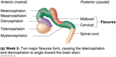
The brain and spinal cord originate from the neural tube during embryonic development. The anterior end of the neural tube expands and forms three primary brain vesicles:
Forebrain (prosencephalon)
Midbrain (mesencephalon)
Hindbrain (rhombencephalon)
The posterior end of the neural tube becomes the spinal cord. As development progresses, these primary vesicles further divide into secondary brain vesicles, giving rise to the major regions of the adult brain.
Brain Regions and Organization
Major Brain Regions
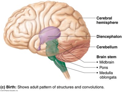
Cerebral Hemispheres
Diencephalon (thalamus, hypothalamus, epithalamus)
Brain Stem (midbrain, pons, medulla oblongata)
Cerebellum
Gray and White Matter
The CNS is organized into gray and white matter:
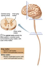
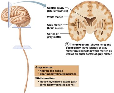
Gray matter: Contains neuron cell bodies, dendrites, and unmyelinated axons. It is the site of synaptic integration and processing.
White matter: Composed mainly of myelinated axons, responsible for transmitting signals between different CNS regions.
The basic pattern is a central cavity surrounded by gray matter, with white matter external to gray matter. This pattern is modified in the brainstem, cerebrum, and cerebellum, where additional gray matter nuclei are present within the white matter, and an outer cortex of gray matter is found in the cerebrum and cerebellum.
Ventricles of the Brain
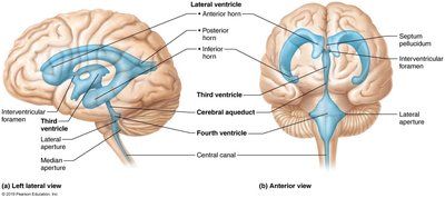
The brain contains fluid-filled chambers called ventricles, which are continuous with each other and the central canal of the spinal cord. They are filled with cerebrospinal fluid (CSF) and lined by ependymal cells. The main ventricles are:
Lateral ventricles: Paired, C-shaped chambers in each hemisphere, separated by the septum pellucidum.
Third ventricle: Located in the diencephalon, connected to the lateral ventricles via the interventricular foramen.
Fourth ventricle: Located in the hindbrain, connected to the third ventricle by the cerebral aqueduct and continuous with the central canal of the spinal cord. It communicates with the subarachnoid space via three openings (lateral and median apertures).
Cerebral Hemispheres
Surface Features and Lobes
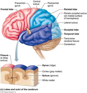
The cerebral hemispheres form the superior part of the brain and account for about 80% of its mass. The surface is marked by:
Gyri: Ridges
Sulci: Shallow grooves
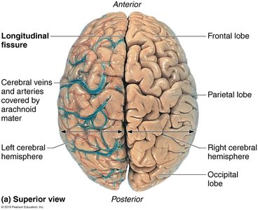
Fissures: Deep grooves (e.g., longitudinal fissure separates hemispheres; transverse cerebral fissure separates cerebrum from cerebellum)
Each hemisphere is divided into five lobes: frontal, parietal, temporal, occipital, and insula (the latter is buried beneath parts of the other lobes).
Functional Areas of the Cerebral Cortex
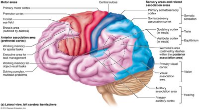
The cerebral cortex is the outer layer of gray matter and is the site of conscious mind functions, including awareness, sensory perception, voluntary motor initiation, communication, memory storage, and understanding. It contains three types of functional areas:
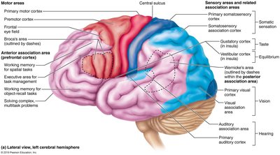
Motor areas: Control voluntary movement (located in the frontal lobe)
Sensory areas: Receive and process sensory information (located in parietal, temporal, occipital, and insular lobes)
Association areas: Integrate information for higher-level processing
Each hemisphere controls the contralateral (opposite) side of the body. There is lateralization of function, with the left hemisphere typically dominant for language, math, and logic, and the right for visual-spatial skills and creativity.
Motor Areas
Primary motor cortex: Located in the precentral gyrus; controls voluntary movements via pyramidal cells and corticospinal tracts.
Premotor cortex: Plans and coordinates learned, repetitive, or patterned motor skills.
Broca's area: Motor speech area, usually in the left hemisphere; involved in speech production.
Frontal eye field: Controls voluntary eye movements.
Sensory Areas
Primary somatosensory cortex: In the postcentral gyrus; receives sensory input from skin and proprioceptors.
Somatosensory association cortex: Integrates sensory input for object recognition.
Visual areas: Primary visual cortex (occipital lobe) and visual association area for interpretation.
Auditory areas: Primary auditory cortex and association area (temporal lobe).
Vestibular cortex: Awareness of balance (insula and parietal cortex).
Olfactory cortex: Smell perception (temporal lobe).
Gustatory cortex: Taste perception (insula).
Visceral sensory area: Perception of internal organ sensations.
Multimodal Association Areas
Anterior association area (prefrontal cortex): Intellect, cognition, personality, working memory, judgment, and planning.
Posterior association area: Pattern and face recognition, spatial localization, language comprehension (Wernicke's area).
Limbic association area: Emotional impact and memory formation (includes hippocampus).
Lateralization and Cerebral Dominance
Lateralization: Specialization of function in each hemisphere.
Cerebral dominance: Hemisphere dominant for language (usually left).
Hemispheres communicate via the corpus callosum and other commissural fibers.
Cerebral White Matter
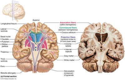
Cerebral white matter is responsible for communication within the cerebrum and between the cerebrum and lower CNS centers. It consists of myelinated fibers organized into tracts:
Association fibers: Connect different parts of the same hemisphere.
Commissural fibers: Connect gray matter of the two hemispheres (e.g., corpus callosum).
Projection fibers: Connect the cerebrum with lower brain regions and the spinal cord.
Basal Nuclei (Ganglia)
The basal nuclei are deep gray matter structures within the cerebral hemispheres. They receive input from the entire cerebral cortex and are closely associated with the diencephalon and midbrain. Their functions include:
Influencing muscle movements and regulating the intensity of slow or stereotyped movements
Playing a role in cognition and emotion
Inhibiting antagonistic or unnecessary movements
The Diencephalon
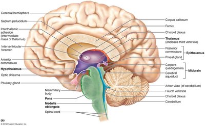
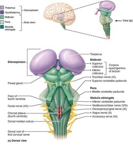
The diencephalon is composed of three gray-matter structures surrounding the third ventricle:
Thalamus: Relay station for sensory and motor signals to the cerebral cortex; sorts, edits, and relays information; involved in sensation, motor activities, learning, and memory.
Hypothalamus: Main visceral control center; regulates autonomic functions (blood pressure, heart rate, digestion, pupil size), emotional responses, body temperature, hunger, thirst, sleep-wake cycles, and endocrine functions (pituitary gland control).
Epithalamus: Contains the pineal gland, which secretes melatonin to regulate sleep-wake cycles.
Clinical Relevance: Homeostatic Imbalances
Damage to specific brain regions can result in loss of function, such as paralysis, sensory deficits, or language impairment. Understanding the anatomical location of functional areas is essential for diagnosing and managing neurological disorders (e.g., stroke, cerebral hemorrhage).
Summary Table: Major Brain Regions and Functions
Region | Main Structures | Primary Functions |
|---|---|---|
Cerebral Hemispheres | Cortex, white matter, basal nuclei | Conscious thought, voluntary movement, sensory perception, memory, language |
Diencephalon | Thalamus, hypothalamus, epithalamus | Relay and processing of sensory information, autonomic and endocrine regulation |
Brain Stem | Midbrain, pons, medulla oblongata | Basic life functions (breathing, heart rate), pathway for tracts, cranial nerves |
Cerebellum | Cerebellar cortex and nuclei | Coordination of movement, balance, motor learning |
