 Indietro
Central Nervous System: Structure and Function of the Brain and Spinal Cord
Guida di studio - Note intelligenti
 Appunti personalizzati basati sui tuoi materiali, ampliati con definizioni chiave, esempi e contesto.
Central Nervous System: The Brain and Spinal Cord
Major Regions of the Brain
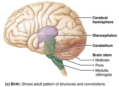
The central nervous system (CNS) consists of the brain and spinal cord, which are responsible for integrating and processing information. The adult brain is divided into four main regions, each with distinct functions:
Cerebral Hemispheres: Responsible for higher cognitive functions, voluntary movement, and sensory perception.
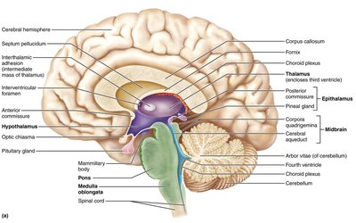
Diencephalon: Contains the thalamus, hypothalamus, and epithalamus, which regulate sensory relay, homeostasis, and endocrine functions.
Brain Stem: Includes the midbrain, pons, and medulla oblongata; controls vital autonomic functions and connects the brain to the spinal cord.
Cerebellum: Coordinates movement and balance.
Basic Structural Patterns in CNS
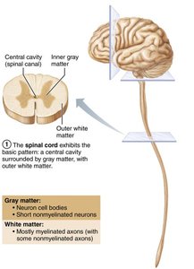
The CNS exhibits a basic pattern of organization with a central cavity surrounded by gray matter, and white matter external to gray matter. This pattern is evident in the spinal cord and changes as it ascends into the brain stem and cerebral hemispheres.
Gray Matter: Composed of neuron cell bodies and short, nonmyelinated neurons.
White Matter: Consists of myelinated and nonmyelinated axons.
Ventricles of the Brain
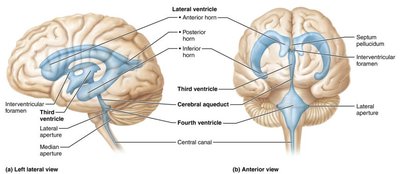
The brain contains fluid-filled chambers called ventricles, which are continuous with each other and the central canal of the spinal cord. These ventricles are filled with cerebrospinal fluid (CSF) and lined by ependymal cells.
Lateral Ventricles: Paired, C-shaped chambers in each hemisphere.
Third Ventricle: Located in the diencephalon, connected to lateral ventricles via interventricular foramen.
Fourth Ventricle: Located between the pons and cerebellum, connected to the third ventricle via the cerebral aqueduct.
Surface Markings and Lobes of the Cerebral Hemispheres
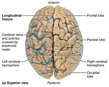
The cerebral hemispheres are marked by ridges (gyri), shallow grooves (sulci), and deep grooves (fissures). These features increase surface area and divide the brain into distinct lobes.
Gyri: Ridges on the brain surface.
Sulci: Shallow grooves separating gyri.
Fissures: Deep grooves, such as the longitudinal fissure (separates hemispheres) and transverse cerebral fissure (separates cerebrum and cerebellum).
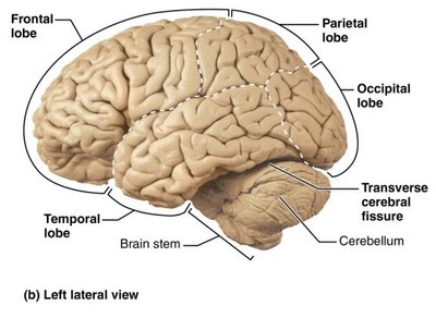
Lobes and Sulci of the Cerebrum
Each hemisphere is divided into five lobes: frontal, parietal, temporal, occipital, and insula. Major sulci separate these lobes and define functional regions.
Central Sulcus: Separates frontal and parietal lobes.
Parieto-occipital Sulcus: Separates parietal and occipital lobes.
Lateral Sulcus: Outlines temporal lobe.
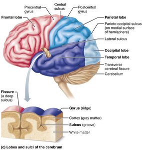
Cerebral Cortex: Functional Areas
The cerebral cortex is the site of conscious mind, including awareness, sensory perception, voluntary motor initiation, communication, memory storage, and understanding. It contains three types of functional areas:
Motor Areas: Control voluntary movement.
Sensory Areas: Provide conscious awareness of sensation.
Association Areas: Integrate diverse information.
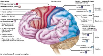
Motor and Sensory Maps
The primary motor cortex and primary somatosensory cortex are organized as maps, with specific regions controlling or sensing different parts of the body. This organization is known as the homunculus.
Motor Map: Located in the precentral gyrus, controls voluntary movements.
Sensory Map: Located in the postcentral gyrus, receives sensory input.
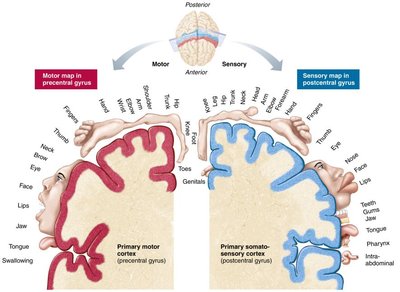
Basal Nuclei and Diencephalon
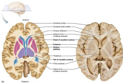
The basal nuclei (ganglia) are deep within the cerebral hemispheres and play a role in regulating movement and cognition. The diencephalon consists of the thalamus, hypothalamus, and epithalamus, which are involved in sensory relay, homeostasis, and endocrine regulation.
Basal Nuclei: Influence muscle movements, filter inappropriate responses, and inhibit unnecessary movements.
Thalamus: Relay station for sensory information.
Hypothalamus: Regulates autonomic functions and endocrine activity.
Epithalamus: Contains the pineal gland, which secretes melatonin.
Brain Stem and Cerebellum
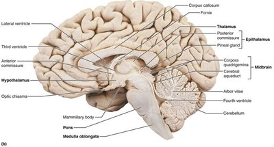
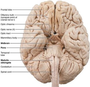
The brain stem connects the brain to the spinal cord and controls vital autonomic functions. The cerebellum coordinates movement and balance.
Midbrain: Connects diencephalon to pons.
Pons: Connects midbrain to medulla oblongata.
Medulla Oblongata: Controls cardiovascular and respiratory functions.
Cerebellum: Processes input for coordinated movement and balance.
Structure of the Spinal Cord
The spinal cord is enclosed in the vertebral column and extends from the foramen magnum to the level of L1 or L2 vertebra. It provides two-way communication between the brain and body and serves as a major reflex center.
Central Canal: Runs the length of the cord and is filled with CSF.
Gray Matter: Contains dorsal, ventral, and lateral horns.
White Matter: Contains ascending, descending, and transverse tracts.
Spinal Tracts and Pathways
Spinal tracts are organized into multineuron pathways that conduct sensory and motor information. Key features include decussation (crossing over), relay (chain of neurons), and symmetry (paired pathways).
Ascending Tracts: Carry sensory information to the brain.
Descending Tracts: Carry motor commands from the brain.
Transverse Tracts: Connect the two sides of the spinal cord.
Meninges: Structure and Function
The meninges are three protective layers that cover the CNS, protect blood vessels, contain CSF, and form partitions within the skull.
Dura Mater: Strongest, outermost layer.
Arachnoid Mater: Middle layer with web-like extensions; contains CSF and blood vessels.
Pia Mater: Delicate, innermost layer that clings to the brain and spinal cord.
Cerebrospinal Fluid: Path and Function
CSF forms a liquid cushion around the brain and spinal cord, providing buoyancy, protection, and nourishment. It is produced by the choroid plexus and circulates through the ventricles, central canal, and subarachnoid space.
Functions: Buoyancy, protection, chemical stability.
Path: Produced in choroid plexus → lateral ventricles → third ventricle → fourth ventricle → subarachnoid space → reabsorbed into venous blood.
Clinical Implications
Stroke (Cerebrovascular Accident): Blockage or rupture of cerebral arteries leads to brain tissue death.
Meningitis: Inflammation of the meninges, diagnosed via CSF analysis.
Hydrocephalus: Obstruction of CSF flow increases intracranial pressure.
Spinal Cord Trauma: Injury can cause sensory or motor loss, depending on location.
Amyotrophic Lateral Sclerosis (ALS): Degeneration of motor neurons in the spinal cord.
Summary Table: Major Regions and Functions of the Brain
Region | Main Function |
|---|---|
Cerebral Hemispheres | Higher cognitive functions, voluntary movement, sensory perception |
Diencephalon | Sensory relay, homeostasis, endocrine regulation |
Brain Stem | Vital autonomic functions, connects brain to spinal cord |
Cerebellum | Coordination of movement and balance |
