 Indietro
Central Nervous System: Structure and Function – Study Notes
Guida di studio - Note intelligenti
 Appunti personalizzati basati sui tuoi materiali, ampliati con definizioni chiave, esempi e contesto.
Central Nervous System (CNS)
Overview of the CNS
The central nervous system (CNS) consists of the brain and spinal cord. It is responsible for integrating sensory information, coordinating bodily functions, and facilitating higher cognitive processes such as thought, memory, and emotion.
Brain: The control center for processing and interpreting sensory information and directing responses.
Spinal Cord: Conducts signals to and from the brain and controls reflex activities.
Directional terms specific to the CNS:
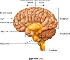
Rostral: Toward the nose or forehead (anterior in the brain).
Caudal: Toward the tail or posterior part of the body.
Organization of Gray and White Matter
Gray Matter
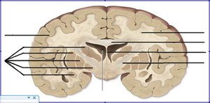
Gray matter consists mainly of neuronal cell bodies, dendrites, and unmyelinated axons. It forms the cortex (outer layer) of the brain and clusters called cerebral nuclei (basal nuclei) deep within the brain.
Cortex: The outermost layer of the cerebrum, responsible for higher-order functions such as perception, thought, and voluntary movement.
Cerebral Nuclei: Clusters of gray matter within the white matter, involved in regulating movement and other functions.
White Matter
White matter is composed of myelinated axons that connect different regions of the CNS. Major structures include:
Corpus Callosum: Connects the left and right cerebral hemispheres, facilitating interhemispheric communication.
Internal Capsule: Contains ascending and descending axons, relaying information between the cortex and lower CNS regions.
Septum Pellucidum: Thin membrane separating the lateral ventricles.
Major Regions of the Brain
Four Major Regions
Cerebrum
Diencephalon
Brainstem
Cerebellum
The cerebrum is the largest region, responsible for higher cognitive functions, voluntary movement, and sensory perception.
Cerebral Cortex and Lobes
Lobes of the Cerebral Cortex
Frontal Lobe: Involved in voluntary movement, planning, reasoning, problem-solving, and speech production.
Parietal Lobe: Processes sensory information such as touch, temperature, and pain.
Temporal Lobe: Responsible for auditory processing, memory, and language comprehension.
Occipital Lobe: Primary center for visual processing.
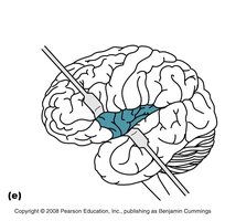
Insula: Involved in taste, visceral sensation, and integrating sensory information.
Gyri and Sulci
Gyri: Elevated ridges on the cerebral surface that increase surface area.
Sulci: Shallow grooves between gyri.
Fissures: Deep grooves that separate major brain regions (e.g., longitudinal and transverse fissures).
The increased surface area provided by gyri and sulci allows for greater cortical processing capacity.
Functional Areas of the Cerebral Cortex
Sensory Areas
Primary Somatosensory Cortex: Located in the postcentral gyrus of the parietal lobe; receives tactile information from the body.
Primary Visual Cortex: Located in the occipital lobe; processes visual information.
Primary Auditory Cortex: Located in the temporal lobe; processes auditory information.
Primary Gustatory Cortex: Located in the insula; processes taste information.
Association Areas
Location: Surround primary sensory and motor areas.
Function: Integrate and interpret sensory input, coordinate responses, and facilitate complex cognitive processes.
Motor Areas
Primary Motor Cortex: Located in the precentral gyrus of the frontal lobe; initiates voluntary movements.
Diencephalon
Thalamus
Location: Deep within the brain, superior to the brainstem.
Structure: Paired, egg-shaped masses of gray matter.
Function: Relay station for sensory information (except olfaction) to the cerebral cortex.
Hypothalamus
Location: Below the thalamus.
Structure: Small region containing multiple nuclei.
Functions: Regulates homeostasis, endocrine activity, autonomic functions, and emotional responses.
Epithalamus (Pineal Gland)
Location: Posterior part of the diencephalon.
Structure: Contains the pineal gland.
Function: Secretes melatonin, regulating circadian rhythms.
Brainstem
Regions and Functions
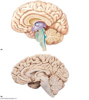
Midbrain: Contains cerebral peduncles (motor tracts), superior and inferior colliculi (visual and auditory reflexes), and cranial nerves III and IV.
Pons: Connects cerebellum to brainstem, relays information, and contains cranial nerves V-VIII.
Medulla Oblongata: Contains pyramids (motor tracts), decussation of fibers, and centers for cardiovascular, respiratory, and reflex functions. Cranial nerves IX-XII attach here.
Cerebellum
Structure and Function
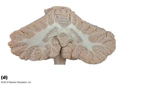
Structure: Two hemispheres connected by the vermis; outer cerebellar cortex (gray matter), inner arbor vitae (white matter), and deep cerebellar nuclei.
Function: Coordinates voluntary movements, maintains posture and balance, and smooths motor activity.
Ventricular System of the Brain
Ventricles and CSF Flow
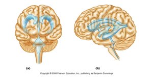
Lateral Ventricles: Located in each cerebral hemisphere; separated by the septum pellucidum.
Third Ventricle: Located in the diencephalon; connected to lateral ventricles by interventricular foramina.
Fourth Ventricle: Located between the pons and cerebellum; continuous with the central canal of the spinal cord.
Choroid Plexus: Produces cerebrospinal fluid (CSF).
CSF flows from the lateral ventricles → third ventricle → cerebral aqueduct → fourth ventricle → subarachnoid space and central canal.
Protection of the CNS
Meninges
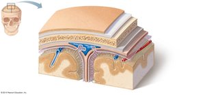
Dura Mater: Tough, outermost layer; has periosteal and meningeal layers in the brain (only meningeal in the spinal cord).
Arachnoid Mater: Middle, web-like layer; subarachnoid space contains CSF and blood vessels.
Pia Mater: Thin, innermost layer; adheres to the surface of the brain and spinal cord.
Cerebrospinal Fluid (CSF)
Location: Fills ventricles, subarachnoid space, and central canal.
Functions: Cushions the CNS, removes waste, and provides nutrients.
Production: By choroid plexuses in the ventricles.
Circulation: Lateral ventricles → third ventricle → fourth ventricle → subarachnoid space/central canal.
Blood-Brain Barrier (BBB)
Function: Protects the brain from harmful substances while allowing passage of essential molecules (e.g., glucose, oxygen).
Structure: Formed by endothelial cells with tight junctions, astrocyte end-feet, and basement membrane.
Exceptions: BBB is reduced or absent in areas such as the choroid plexus, hypothalamus, and pineal gland to allow for hormone release and monitoring of blood composition.
Functional Brain Systems
Limbic System
Amygdala: Emotion processing, especially fear and aggression.
Hippocampus: Memory formation and spatial navigation.
Cingulate Gyrus: Emotional regulation and pain processing.
Reticular Formation and Reticular Activating System (RAS)
Location: Extends through the brainstem.
Function: Regulates arousal, consciousness, and sleep-wake cycles.
Levels of Consciousness: Alertness, drowsiness, stupor, coma.
Higher Order Functions
Sleep
NREM (Non-Rapid Eye Movement) Sleep: Deep, restorative sleep with slow brain waves.
REM (Rapid Eye Movement) Sleep: Dreaming occurs, brain activity resembles wakefulness, but muscles are relaxed.
Memory
Memory: The ability to store and retrieve information.
Memory Trace: Physical change in the brain representing memory.
Short-term Memory: Temporary storage of information.
Long-term Memory: More permanent storage, requiring consolidation.
Consolidation: Process of converting short-term to long-term memory.
Brain Areas: Hippocampus (declarative memory), amygdala (emotional memory), cerebellum (procedural memory).
Spinal Cord
Structure and Regions
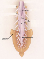
Begins: At the foramen magnum of the skull.
Ends: At the conus medullaris (around L1-L2 vertebrae).
Associated Structures: Conus medullaris (tapered end), filum terminale (fibrous extension anchoring cord), cauda equina (bundle of spinal nerves below conus).
Gray and White Matter in the Spinal Cord
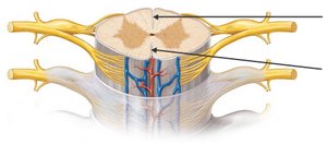
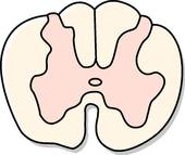
Gray Matter: Located centrally in an H-shaped (butterfly) pattern; contains cell bodies and interneurons.
Regions: Dorsal (posterior) horns (sensory), ventral (anterior) horns (motor), lateral horns (autonomic motor).
White Matter: Surrounds gray matter; contains ascending (sensory) and descending (motor) tracts, organized into columns (funiculi): dorsal, lateral, and ventral.
Spinal Nerves and Segments
Spinal Nerves: 31 pairs; part of the peripheral nervous system (PNS).
Spinal Segments: Defined by the origin of spinal nerves; used clinically to localize lesions or injuries.
Functions of the Spinal Cord
Conducts sensory and motor information between the body and brain.
Coordinates reflexes.
Serves as a center for pattern generators (e.g., walking).
Protection of the Spinal Cord
Vertebrae: Bony protection.
Meninges: Dura mater (single meningeal layer in spinal cord), arachnoid mater, pia mater.
Cerebrospinal Fluid: Cushions and nourishes the spinal cord.
Epidural Space: Located between dura mater and vertebral wall; contains fat and blood vessels.
