 Indietro
Central Nervous System: Structure and Protection of the Brain
Guida di studio - Note intelligenti
 Appunti personalizzati basati sui tuoi materiali, ampliati con definizioni chiave, esempi e contesto.
Brain Regions and Organization
Major Regions of the Adult Brain
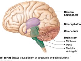
The adult human brain is organized into four primary regions, each with distinct functions and anatomical features. Understanding these regions is essential for comprehending the central nervous system's structure and function.
Cerebral Hemispheres: The largest portion, responsible for higher cognitive functions.
Diencephalon: Contains key relay and regulatory centers.
Brain Stem: Includes the midbrain, pons, and medulla oblongata; controls vital functions.
Cerebellum: Coordinates movement and balance.
Structural Overview
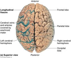
Cerebral Hemispheres: Divided into left and right hemispheres by the longitudinal fissure.
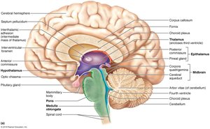
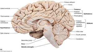
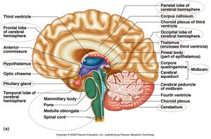
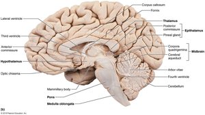
Diencephalon: Located centrally, includes thalamus, hypothalamus, and epithalamus.
Brain Stem: Connects the brain to the spinal cord; essential for autonomic functions.
Cerebellum: Sits posterior to the brain stem, involved in motor control.
Ventricles of the Brain
Structure and Function
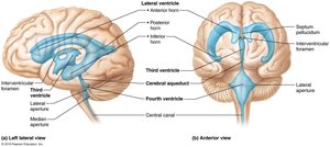
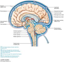
The brain contains interconnected, fluid-filled chambers called ventricles. These are essential for the circulation of cerebrospinal fluid (CSF), which cushions and nourishes the brain.
Lateral Ventricles: Paired, located in each cerebral hemisphere; separated by the septum pellucidum.
Third Ventricle: Located in the diencephalon; connected to lateral ventricles via interventricular foramen.
Fourth Ventricle: Located in the hindbrain; connected to the third ventricle via the cerebral aqueduct.
CSF Flow: CSF circulates through ventricles and into the subarachnoid space, providing protection and nutrient transport.
Cerebral Hemispheres
Surface Markings and Lobes
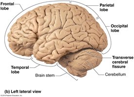
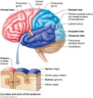
The cerebral hemispheres are characterized by distinct surface features and are divided into lobes, each associated with specific functions.
Gyri: Ridges on the brain surface.
Sulci: Shallow grooves separating gyri.
Fissures: Deep grooves, such as the longitudinal and transverse cerebral fissures.
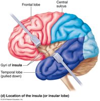
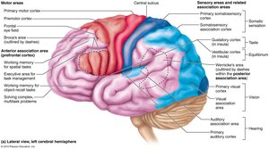
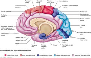
Lobes: Frontal, parietal, temporal, occipital, and insula (buried beneath other lobes).
Functional Areas
Motor Areas: Located in the frontal lobe; control voluntary movements.
Sensory Areas: Located in parietal, occipital, and temporal lobes; process sensory information.
Association Areas: Integrate information for complex functions such as reasoning and memory.
Diencephalon
Components and Functions
The diencephalon is a central brain region composed of three paired gray-matter structures:
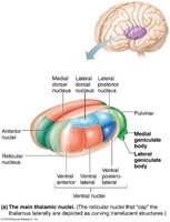
Thalamus: Acts as a relay station for sensory and motor signals to the cerebral cortex.
Hypothalamus: Regulates homeostasis, including temperature, hunger, thirst, and endocrine functions.
Epithalamus: Contains the pineal gland, which secretes melatonin to regulate sleep-wake cycles.
Protection of the Brain
Meninges
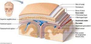
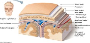
The brain is protected by three connective tissue layers called meninges. These layers provide structural support, protect blood vessels, and contain cerebrospinal fluid.
Pia Mater: Innermost, delicate layer that closely follows brain contours; rich in blood vessels.
Arachnoid Mater: Middle layer with web-like extensions; separated from pia mater by the subarachnoid space, which contains CSF.
Dura Mater: Outermost, tough layer; consists of periosteal and meningeal layers, forms partitions (dural septa) within the skull.
Dural Septa and Sinuses
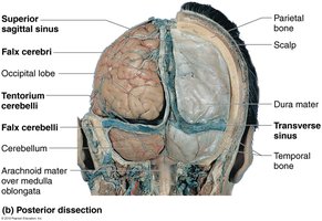
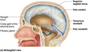
Falx Cerebri: Partition in the longitudinal fissure.
Falx Cerebelli: Partition along the vermis of the cerebellum.
Tentorium Cerebelli: Horizontal fold over the cerebellum.
Dural Venous Sinuses: Collect venous blood and drain into jugular veins.
Cerebrospinal Fluid (CSF)
Formation and Function
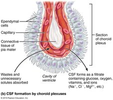
CSF: A clear, colorless fluid that cushions the brain, reduces its weight, and provides nutrients.
Choroid Plexus: Specialized capillary network in ventricles that produces CSF.
CSF Circulation: Flows through ventricles, subarachnoid space, and is reabsorbed into venous blood via arachnoid granulations.
Sheep Brain Dissection
Comparative Anatomy
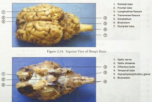
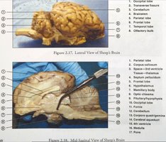
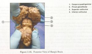
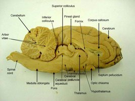
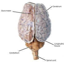
Sheep brain dissections are commonly used in laboratory settings to illustrate the anatomy of the central nervous system. Key structures are similar to those in the human brain, making it a valuable model for study.
Major Structures: Cerebrum, cerebellum, brainstem, optic chiasma, pineal gland, corpus callosum, and ventricles.
Orientation: Superior, lateral, and midsagittal views help identify anatomical landmarks.
Summary Table: Major Brain Regions and Functions
Region | Main Structures | Primary Functions |
|---|---|---|
Cerebral Hemispheres | Frontal, parietal, temporal, occipital, insula | Conscious thought, voluntary movement, sensory processing |
Diencephalon | Thalamus, hypothalamus, epithalamus | Relay, regulation, homeostasis, endocrine control |
Brain Stem | Midbrain, pons, medulla oblongata | Autonomic functions, cranial nerve nuclei, fiber tracts |
Cerebellum | Anterior, posterior, flocculonodular lobes | Coordination, balance, motor learning |
Key Equations and Concepts
CSF Volume Replacement
Normal adult CSF volume: ~150 ml
Replaced every 8 hours
Example Equation: CSF Turnover Rate
Let V = CSF volume, T = time for replacement
Clinical Note
Meningitis and Encephalitis
Meningitis: Inflammation of the meninges, diagnosed via CSF analysis.
Encephalitis: Inflammation of the brain, often secondary to meningitis.
Additional info:
Sheep brain models are used to reinforce anatomical concepts and provide hands-on learning for students.
CSF is essential for maintaining the brain's environment and protecting it from injury.
