 Indietro
Central Nervous System: Structure, Function, and Protection
Guida di studio - Note intelligenti
 Appunti personalizzati basati sui tuoi materiali, ampliati con definizioni chiave, esempi e contesto.
Central Nervous System Overview
Divisions of the Nervous System
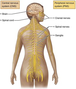
The nervous system is divided into two principal parts: the Central Nervous System (CNS) and the Peripheral Nervous System (PNS). The CNS consists of the brain and spinal cord, serving as the integration and control center. The PNS includes nerves and ganglia outside the CNS, connecting the CNS to limbs and organs.
CNS: Brain and spinal cord; interprets sensory input and dictates motor output.
PNS: Cranial nerves, spinal nerves, ganglia; transmits signals between CNS and the rest of the body.
Somatic vs. Autonomic Pathways
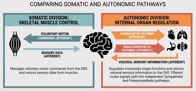
The nervous system is further divided into somatic and autonomic pathways. The somatic division manages voluntary motor commands and sensory data from muscles, while the autonomic division regulates involuntary organ functions through sympathetic and parasympathetic pathways.
Somatic Division: Controls skeletal muscle movement; relays sensory information from muscles.
Autonomic Division: Controls internal organ regulation; includes sympathetic (fight or flight) and parasympathetic (rest and digest) pathways.
Structural Organization of the CNS
Gray Matter and White Matter
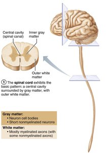
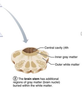
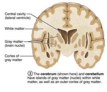
The CNS is organized into regions of gray matter and white matter. Gray matter consists of neuron cell bodies and unmyelinated neurons, while white matter is composed of myelinated axons.
Spinal Cord: Central cavity surrounded by gray matter, with outer white matter.
Brain Stem: Central cavity (4th ventricle), inner gray matter, outer white matter, and additional gray matter nuclei.
Cerebrum and Cerebellum: Islands of gray matter (nuclei) within white matter, plus an outer cortex of gray matter.
Ventricles of the Brain
Structure and Function
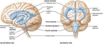
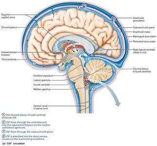
The brain contains fluid-filled chambers called ventricles, which are continuous with each other and the central canal of the spinal cord. They are filled with cerebrospinal fluid (CSF) and lined by ependymal cells.
Lateral Ventricles: Large, C-shaped chambers in each hemisphere.
Third Ventricle: Located in the diencephalon.
Fourth Ventricle: Located in the hindbrain, continuous with the central canal.
CSF: Provides buoyancy, protection, and nourishment to the brain.
Protection of the Brain
Meninges
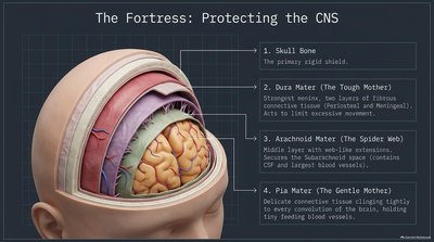
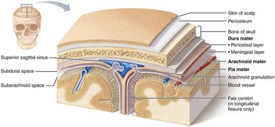
The brain is protected by three connective tissue layers called meninges: dura mater, arachnoid mater, and pia mater. These layers cover and protect the CNS, enclose blood vessels, and contain CSF.
Dura Mater: Tough, outermost layer; limits excessive movement.
Arachnoid Mater: Middle layer with web-like extensions; contains CSF and blood vessels.
Pia Mater: Delicate, innermost layer; clings tightly to the brain and contains blood vessels.
Cerebrospinal Fluid (CSF)
Formation, Circulation, and Function
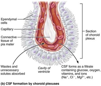
Cerebrospinal fluid (CSF) forms a liquid cushion around the brain, providing buoyancy, protection, and nourishment. It is produced by the choroid plexus in each ventricle and circulates through the ventricles and subarachnoid space, eventually being absorbed into the dural venous sinuses.
Formation: CSF is formed from blood plasma by the choroid plexus.
Circulation: Flows through ventricles, subarachnoid space, and is absorbed via arachnoid granulations.
Function: Reduces brain weight, protects from trauma, and carries chemical signals.
Blood Brain Barrier
Structure and Function
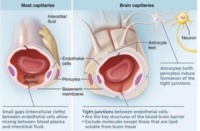
The blood brain barrier maintains a stable environment for the brain by regulating the passage of substances from the blood into the CNS. It consists of tight junctions between endothelial cells, supported by astrocyte feet and pericytes.
Tight Junctions: Prevent substances from passing between cells.
Astrocytes and Pericytes: Promote tight-junction formation.
Transport Mechanisms: Simple diffusion, facilitated diffusion, and transcytosis.
Basement Membrane: Contains enzymes that destroy chemicals harmful to neurons.
Major Brain Regions and Their Functions
Cerebrum, Diencephalon, Brain Stem, Cerebellum
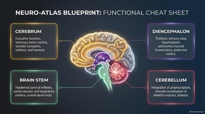
The brain is divided into four major regions, each with distinct functions:
Cerebrum: Executive function, voluntary motor control, somatic sensation, intellect, and memory.
Diencephalon: Thalamic sensory relay, hypothalamic autonomic/visceral homeostasis, endocrine control.
Brain Stem: Survival reflexes, cardiovascular and respiratory centers, cranial nerve roots.
Cerebellum: Integration of proprioception, smooth coordination of skeletal muscles, balance.
White Fiber Tracts of the Cerebral Hemispheres
Types of Fiber Tracts
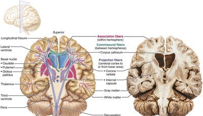
White matter in the cerebral hemispheres is organized into three types of fiber tracts:
Association Fibers: Connect different parts of the same hemisphere.
Commissural Fibers: Connect gray matter of the two hemispheres (e.g., corpus callosum).
Projection Fibers: Connect the hemispheres with lower brain or spinal cord.
Spinal Cord Anatomy
Cross-Sectional Architecture
The spinal cord is enclosed in the vertebral column and consists of a central core of gray matter surrounded by white matter. It provides two-way communication between the brain and body and serves as a major reflex center.
Gray Matter: Contains nerve cell bodies; forms the central butterfly or "H" shape.
White Matter: Contains myelinated nerve tracts; organized into dorsal, ventral, and lateral columns.
Central Canal: Runs the length of the cord and is filled with CSF.
Gross Structure and Protection
The spinal cord is protected by bone, meninges, and CSF. The dura mater is a single layer and does not attach to the vertebrae. The epidural space contains fat and veins, while the subarachnoid space is filled with CSF.
Cervical and Lumbar Enlargements: Where nerves serving the limbs emerge.
Conus Medullaris: Tapered inferior end of the spinal cord.
Cauda Equina: Bundle of nerve roots below the conus medullaris.
Filum Terminale: Fibrous extension anchoring the cord to the coccyx.
Spinal Cord Segments
Spinal cord segments are aligned with their vertebrae in the cervical region, but in the lumbar and sacral regions, segments are not adjacent to their corresponding vertebrae due to differential growth rates during development.
Cervical Segments: C1–C7
Thoracic Segments: T1–T12
Lumbar and Sacral Segments: Not adjacent to corresponding vertebrae
Lumbar Puncture
A lumbar puncture is performed by inserting a needle between the L4 and L5 vertebrae to access the subarachnoid space for CSF sampling.
Needle Path: Passes through ligaments and enters the subarachnoid space.
Structures Shown: Cauda equina, spinal dura mater, arachnoid mater, intervertebral discs.
Gray Matter and Spinal Roots
Each side of the spinal cord contains three areas of gray matter: dorsal horns (sensory input), ventral horns (motor output), and lateral horns (sympathetic neurons).
Dorsal Horns: Receive somatic and visceral sensory input.
Ventral Horns: Contain somatic motor neurons.
Lateral Horns: Present in thoracic and superior lumbar regions; contain sympathetic neurons.
Neural Routing: Sensory and Motor Pathways
Sensory information enters the spinal cord via the dorsal root ganglion and dorsal horn, while motor commands exit via the ventral horn and ventral root. The spinal nerve acts as a two-way conduit before splitting into distinct roots.
Sensory Pathways: Peripheral receptors → dorsal root ganglion → dorsal root → dorsal horn.
Motor Pathways: Ventral horn → ventral root → peripheral effectors.
Organization of Gray Matter
The gray matter of the spinal cord is organized into four functional zones: somatic sensory (SS), visceral sensory (VS), visceral motor (VM), and somatic motor (SM).
SS: Somatic sensory
VS: Visceral sensory
VM: Visceral motor
SM: Somatic motor
----------------------------------------
