 Indietro
Central Nervous System Study Guide – Anatomy & Physiology
Guida di studio - Note intelligenti
 Appunti personalizzati basati sui tuoi materiali, ampliati con definizioni chiave, esempi e contesto.
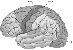
Q10. Select the letter that represents the location of the primary motor cortex.
Background
Topic: Brain Anatomy – Functional Areas
This question tests your knowledge of the anatomical location of the primary motor cortex in the cerebrum, which is responsible for voluntary motor control.
Key Terms:
Primary motor cortex: Region of the frontal lobe responsible for initiating voluntary movements.
Precentral gyrus: The specific gyrus where the primary motor cortex is located.
Step-by-Step Guidance
Examine the labeled regions (A, B, C, D) on the provided brain diagram.
Recall that the primary motor cortex is found in the precentral gyrus of the frontal lobe.
Identify which labeled region corresponds to the precentral gyrus.
Compare the location of each label with the anatomical landmarks of the frontal lobe.
Try solving on your own before revealing the answer!
Final Answer: A
The primary motor cortex is located at label A, which corresponds to the precentral gyrus of the frontal lobe.
This area is responsible for voluntary motor control of skeletal muscles.
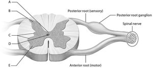
Q: Match the following with the spinal cord cross section.
Background
Topic: Spinal Cord Anatomy
This question tests your ability to identify key anatomical structures in a cross-section of the spinal cord, including gray and white matter regions and their functions.
Key Terms:
Gray commissure: Thin strip of gray matter surrounding the central canal.
Lateral horn: Contains cell bodies for autonomic motor control.
Posterior horn: Processes somatic and visceral sensory information.
Step-by-Step Guidance
Review the labeled parts (A-E) on the spinal cord cross-section diagram.
Recall the functions of the anterior, posterior, and lateral horns of gray matter.
Identify the gray commissure (thin strip around the central canal).
Locate the lateral horn (small lateral projection between the horns).
Determine which region contains cell bodies for sensory processing (posterior horn).
Try solving on your own before revealing the answer!
Final Answers:
The structure that contains cell bodies for motor control of viscera via the autonomic nervous system: C (lateral horn).
The gray commissure: A.
Cell bodies for processing somatic and visceral incoming sensory information: B (posterior horn).
These regions are critical for the integration and relay of motor and sensory signals in the spinal cord.
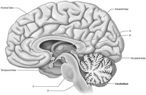
Q: Match the following with the sagittal section of the brain.
Background
Topic: Brain Anatomy – Sagittal Section
This question tests your ability to identify major structures in a sagittal section of the brain and understand their functions.
Key Terms:
Hypothalamus: Regulates body temperature, sleep/wake cycle, hunger, and thirst.
Pons: Part of the brainstem involved in relaying information and regulating vital functions.
Thalamus: Edits, sorts, and routes stimuli; controls entry of information into the cerebral cortex.
Step-by-Step Guidance
Examine the labeled parts (A-D) on the sagittal brain diagram.
Recall the location and function of the hypothalamus, pons, and thalamus.
Identify the hypothalamus (small area below the thalamus, near the center).
Locate the pons (stem-like structure below the cerebellum).
Determine which structure edits, sorts, and routes stimuli (egg-shaped structure in the middle).
Try solving on your own before revealing the answer!
Final Answers:
Structure that regulates body temperature, sleep/wake cycle, hunger, and thirst: B (hypothalamus).
Pons: D.
Structure that edits, sorts, and routes stimuli and controls entry into the cerebral cortex: A (thalamus).
These structures are essential for homeostasis, sensory relay, and integration of information in the brain.
