 Indietro
E2 Lecture 5: Central Nervous System (CNS)
Guida di studio - Note intelligenti
 Appunti personalizzati basati sui tuoi materiali, ampliati con definizioni chiave, esempi e contesto.
Central Nervous System
The Brain and Spinal Cord: Overview
The central nervous system (CNS) consists of the brain and spinal cord, which are responsible for integrating sensory information and coordinating bodily functions. The spinal cord is enclosed within the vertebral column, beginning at the foramen magnum and ending at the L1 or L2 vertebra. It serves as a major pathway for communication between the brain and the rest of the body and is the site for reflex actions.
Two-way communication: The spinal cord transmits sensory signals to the brain and motor signals to muscles and glands.
Reflex center: Reflexes are initiated and completed within the spinal cord.
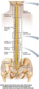
Spinal Cord: Functional Anatomy
The spinal cord contains bundles of axons that conduct information up and down its length. Sensory signals move from the body to the brain, while motor signals travel from the brain to muscles and glands below the head.
Conduction: Bundles of axons transmit information.
Sensory and motor pathways: Ascending tracts carry sensory information; descending tracts carry motor commands.
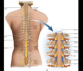
Gross Anatomy and Protection
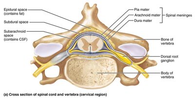
The spinal cord is protected by bone, meninges, and cerebrospinal fluid (CSF). The meninges are three protective membranes: dura mater (outermost), arachnoid mater (middle), and pia mater (innermost). The CSF fills the subarachnoid space between the arachnoid and pia maters, providing cushioning and chemical stability.
Spinal dura mater: Tough, outer membrane.
Epidural space: Space containing fat and blood vessels.
CSF: Fills subarachnoid space, cushions and nourishes the CNS.
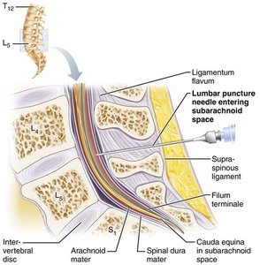
Site of lumbar puncture: Dural and arachnoid membranes extend to the sacrum, allowing safe access for CSF sampling.
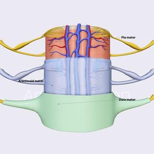
Spinal Meninges – Protective Membranes
The brain and spinal cord are covered by three membranes that prevent abrasion and trauma. These are:
Dura mater: Outermost, tough membrane against bone.
Arachnoid mater: Middle, web-like layer.
Pia mater: Innermost, delicate layer adhering to nervous tissue.
Gross Anatomy: Conus Medullaris, Filum Terminale, and Cauda Equina
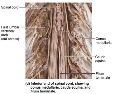
The spinal cord terminates in a cone-shaped structure called the conus medullaris. The filum terminale is a fibrous extension of the pia mater that anchors the cord to the coccyx. The cauda equina is a bundle of spinal nerves and nerve roots below the conus medullaris.
Denticulate ligaments: Extensions of pia mater that secure the cord to dura mater.
Cervical and lumbar enlargements: Regions where nerves serving upper and lower limbs arise.
Spinal Nerves and Segmentation
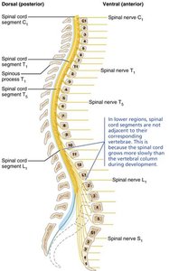
Spinal nerves are part of the peripheral nervous system (PNS) and attach to the spinal cord by 31 paired roots. Each segment of the spinal cord is designated by the paired spinal nerves that arise from it. The cauda equina consists of nerve roots that extend below the spinal cord.
Cervical and lumbosacral enlargements: Nerves serving upper and lower limbs emerge here.
Segmentation: Each segment corresponds to a pair of spinal nerves.
Spinal Cord Cross-Sectional Anatomy
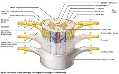
In cross-section, the spinal cord has gray matter in the core and white matter outside. The central canal runs the length of the cord and is filled with CSF.
Gray matter: Contains neuron cell bodies, dendrites, and glial cells.
White matter: Contains myelinated axons organized into tracts.
Central canal: Filled with CSF, runs longitudinally.
Spinal Cord Trauma and Disorders
Functional Losses Due to Injury
Localized injury to the spinal cord or its roots can lead to sensory or motor function loss. Paresthesia is caused by damage to dorsal roots or sensory tracts, resulting in sensory loss. Paralysis is caused by damage to ventral roots or ventral horn cells, resulting in motor loss.
Flaccid paralysis: Severe damage; impulses do not reach muscles; no voluntary or involuntary control; muscles atrophy.
Spastic paralysis: Damage to upper motor neurons; muscles are stimulated by reflex activity; no voluntary control; muscles may shorten permanently.
Types of Spinal Cord Trauma
Transection: Cross-sectioning of the spinal cord results in total motor and sensory loss below the cut.
Paraplegia: Transection between T1 and L1.
Quadriplegia: Transection in the cervical region.
Spinal shock: Temporary loss of function below the lesion.
Amyotrophic Lateral Sclerosis (ALS)
ALS, also known as Lou Gehrig’s disease, involves destruction of ventral horn motor neurons and fibers of the pyramidal tract. Symptoms include loss of ability to speak, swallow, and breathe. Death typically occurs within five years. Causes include environmental factors and genetic mutations affecting RNA processing.
Brain: Structure and Functional Regions
Meninges of the Brain
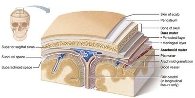
The brain is protected by three meninges: dura mater (thickest, forms fibrous sac and dural sinuses), arachnoid mater (contains large blood vessels in subarachnoid space), and pia mater (thinnest, inner layer).
Cerebral Hemispheres
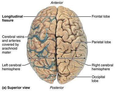
The cerebral hemispheres form the superior part of the brain and account for 83% of its mass. Surface markings include gyri (ridges), sulci (shallow grooves), and fissures (deep grooves). The longitudinal fissure separates the two hemispheres.
Lobes of the Cerebral Hemispheres
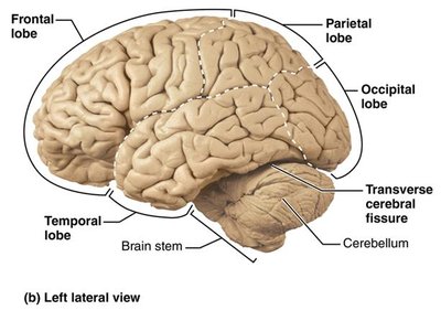
Each hemisphere is divided into five lobes: frontal, parietal, temporal, occipital, and insula (buried under portions of temporal, parietal, and frontal lobes).
Basic Regions of the Cerebral Hemispheres
Cerebral cortex: Superficial layer of gray matter; site of conscious mind, awareness, sensory perception, voluntary motor initiation, communication, memory, and understanding.
Internal white matter: Myelinated axons connecting different regions.
Basal nuclei: Deep within white matter; influence muscle movements and play roles in cognition and emotion.
Cerebral Cortex: Functional Areas
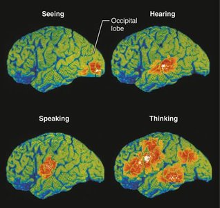
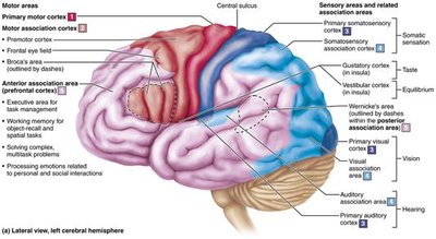
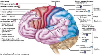
Motor areas: Control voluntary movement; located in frontal lobe.
Sensory areas: Conscious awareness of sensation; located in parietal, insular, temporal, and occipital lobes.
Association areas: Integrate diverse information; multimodal association areas receive inputs from multiple sensory areas and send outputs to multiple areas.
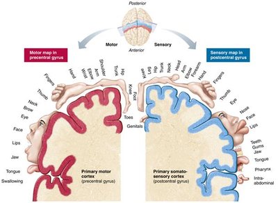
Motor Areas of the Cerebral Cortex
Primary motor cortex: Controls voluntary movements.
Premotor cortex: Plans movements and coordinates learned motor skills.
Frontal eye field: Controls voluntary eye movements.
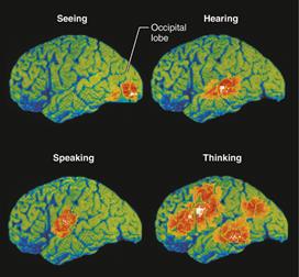
Sensory Areas of the Cerebral Cortex
Primary somatosensory cortex: Receives general sensory information from skin, skeletal muscle, joints, and tendons.
Visual cortex: Interprets visual stimuli.
Auditory and olfactory cortex: Processes sound and smell.
Association Areas of the Cerebral Cortex
Multimodal association areas: Integrate information from multiple sensory modalities, store memories, and tie to previous experiences.
Conscious behavior: Involves the entire cortex.
Cerebral White Matter
Tracts: Bundles of myelinated axons connecting areas of the CNS.
Corpus callosum: Largest tract connecting the two hemispheres.
Basal Nuclei (Basal Ganglia)
Muscle movement regulation: Overlap with cerebellum; role in cognition and emotion.
Movement intensity: Regulate slow or stereotyped movements.
Filtering: Filter out incorrect or inappropriate responses.
Inhibition: Inhibit antagonistic or unnecessary movements.
Divisions of the Brain
Cerebral hemispheres
Diencephalon: Thalamus, hypothalamus, pineal gland
Brain stem: Midbrain, pons, medulla oblongata
Cerebellum
Diencephalon
Thalamus: Relay station for information entering the cortex; sorts, edits, and relays ascending input.
Hypothalamus: Controls autonomic nervous system, initiates physical responses to emotions, regulates body temperature, hunger, thirst, sleep-wake cycles, and endocrine function.
Pineal gland: Secretes melatonin, regulates sleep-wake cycles.
Brain Stem
Midbrain: Filters pain stimuli, produces dopamine, acts on basal nuclei, involved in vision and hearing.
Pons: Connects higher brain centers and spinal cord, relays impulses between motor cortex and cerebellum, origin of cranial nerves V, VI, VII.
Medulla oblongata: Autonomic reflex center; cardiovascular, vasomotor, and respiratory centers; controls vomiting, swallowing, coughing, sneezing.
Cerebellum
Coordination: Processes input to provide precise, coordinated movements of skeletal muscles.
Balance: Plays a major role in balance and timing.
Functional Brain Systems
Limbic System
Emotion: Puts emotional responses to odors; interacts with prefrontal lobes for conscious emotional awareness.
Memory: Hippocampus and amygdala coordinate memory formation and emotional significance.
Reticular Formation
Reticular activating system (RAS): Keeps cortex conscious and alert; filters out repetitive or weak stimuli.
Visceral motor functions: Regulates autonomic functions.
Cerebrospinal Fluid (CSF) and Ventricles
Cerebrospinal Fluid (CSF)
Production: Produced by ependymal cells lining ventricles.
Functions: Provides chemical environment, removes wastes, provides buoyancy, cushions the brain.
Ventricles
Structure: Fluid-filled chambers continuous with central canal of spinal cord.
Lined by ependymal cells: Paired lateral ventricles are large, C-shaped chambers deep in each hemisphere.
Blood Supply and Blood Brain Barrier
Blood Brain Barrier
Function: Maintains stable environment for the brain.
Structure: Substances must pass through continuous endothelium of capillary wall; tight junctions ensure selective permeability.
Astrocytes: Promote tight junction formation.
Transport Mechanisms
Simple diffusion: Lipid-soluble substances and blood gases pass freely.
Facilitated diffusion: Moves glucose, amino acids, and specific ions.
Barrier absence: Some areas, such as brain stem and hypothalamus, lack the barrier.
Summary Table: Spinal Cord and Brain Protection
Structure | Function |
|---|---|
Dura mater | Outer protective membrane; forms dural sinuses |
Arachnoid mater | Middle membrane; contains blood vessels |
Pia mater | Inner membrane; adheres to nervous tissue |
CSF | Cushions, nourishes, and removes wastes |
Blood brain barrier | Maintains chemical environment; selective permeability |
Clinical Considerations
Spinal Cord Injuries
Serious consequences: Disrupt communication between brain and body, leading to paralysis and loss of function.
Temporary vs. permanent: Some injuries cause temporary paralysis; others result in permanent loss.
ALS (Amyotrophic Lateral Sclerosis)
Motor neuron destruction: Loss of ability to speak, swallow, and breathe.
Rapid progression: Death typically within five years.
Participation Questions
Why are spinal cord injuries so serious? C. They can disrupt communication between the brain and the body, leading to paralysis and loss of function.
Why do scents trigger strong, vivid memories? D. All of the above: Scents bypass the thalamus, olfactory bulb is close to amygdala and hippocampus, and has direct connections to these regions crucial for emotions and memory.
