 Indietro
Chapter 10: Muscle Tissue - Structure, Function, and Physiology
Guida di studio - Note intelligenti
 Appunti personalizzati basati sui tuoi materiali, ampliati con definizioni chiave, esempi e contesto.
Muscle Tissue Overview
Types of Muscle Tissue
Muscle tissue is a primary tissue type in the human body, classified into three distinct types based on structure and function:
Skeletal muscle: Voluntary, striated, long fibers, multiple nuclei. Responsible for movement of the skeleton.
Cardiac muscle: Involuntary, striated, branched cells, single nucleus, contains intercalated disks. Found only in the heart.
Smooth muscle: Involuntary, non-striated, spindle-shaped cells, single nucleus. Located in walls of hollow organs.
Functions of the Muscular System
Produce movement: Muscles contract and pull on tendons to move bones.
Maintain posture and body position: Muscle tension stabilizes posture.
Support soft tissues: Muscles form body walls and support organs.
Guard entrances and exits: Sphincter muscles control openings of digestive and urinary tracts.
Maintain body temperature: Muscle activity generates heat.
Store nutrients: Muscles store glycogen and proteins.
Organization and Structure of Skeletal Muscle
Muscle Organization
Skeletal muscles are organized hierarchically:
Whole muscle → Fascicles → Muscle fiber → Myofibril → Sarcomere → Myofilament
Three layers of connective tissue surround muscles:
Epimysium: Surrounds the entire muscle.
Perimysium: Surrounds fascicles.
Endomysium: Surrounds individual muscle fibers.
Tendons attach muscles to bones, formed by the merging of connective tissue layers.
Muscle Attachment
Origin: Attachment point that does not move during contraction.
Insertion: Attachment point that moves during contraction.
Action: The movement produced by muscle contraction.
Muscle belly: Central, bulging part of the muscle.
Skeletal Muscle Fiber Structure
Features of Muscle Fibers
Large, multinucleate cells formed by fusion of myoblasts.
Satellite cells: Myoblasts that do not fuse, involved in repair.
Muscle Fiber Components
Sarcolemma: Cell membrane of muscle fiber, conducts electrical impulses.
Sarcoplasm: Cytoplasm of muscle fiber.
Transverse tubules (T-tubules): Extensions of sarcolemma, transmit impulses deep into fiber.
Myofibrils and Myofilaments
Myofibrils: Bundles of myofilaments responsible for contraction.
Myofilaments: Two types—thin (actin) and thick (myosin).
Sarcoplasmic Reticulum (SR)
Specialized smooth ER in muscle fibers.
Forms terminal cisternae, stores and releases Ca2+.
Triad: 1 T-tubule + 2 terminal cisternae.
Sarcomere Structure and Function
Sarcomere Components
Thick filaments: Myosin protein.
Thin filaments: Actin protein.
Titin: Elastic protein, stabilizes filaments.
Troponin and Tropomyosin: Regulate contraction.
Parts of a Sarcomere
A band: Contains thick and thin filaments.
M line: Center of A band, connects thick filaments.
H band: Only thick filaments.
Zone of overlap: Thick and thin filaments overlap.
I band: Only thin filaments.
Z lines: Boundary of sarcomere.
Thin Filament Structure
F actin: Two twisted strands of G actin.
Nebulin: Holds F actin together.
Tropomyosin: Covers active sites on G actin.
Troponin: Three subunits, binds to tropomyosin, G actin, and Ca2+.
Thick Filament Structure
Myosin tails: Form thick filament backbone.
Myosin heads: Bind to actin, form cross-bridges, powered by ATP.
Muscle Contraction Mechanism
Sliding Filament Theory
Muscle contraction occurs as thin filaments slide past thick filaments, shortening the sarcomere:
H and I bands shrink.
Zone of overlap increases.
Z lines move closer together.
A band remains unchanged.
Neuromuscular Junction (NMJ)
Synapse between motor neuron and muscle fiber.
Neuron releases acetylcholine (ACh) into synaptic cleft.
ACh binds to nicotinic acetylcholine receptors on sarcolemma, causing Na+ influx and depolarization.
ACh is broken down by acetylcholinesterase (AChE).
Excitation-Contraction Coupling
Action potential travels along sarcolemma and T-tubules.
Triggers Ca2+ release from SR.
Ca2+ binds to troponin, moves tropomyosin, exposes actin active sites.
Myosin heads bind to actin, forming cross-bridges.
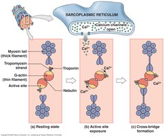
Steps in Muscle Contraction
Ca2+ influx exposes actin active sites.
Myosin cross-bridge formation.
Power stroke: myosin head moves actin filament, powered by ATP.
ATP binds to myosin, detaches cross-bridge.
ATP hydrolysis re-energizes myosin.
Ca2+ pumped back to SR, actin sites covered, muscle relaxes.
Muscle Relaxation
Requires AChE activity, ATP binding to myosin, and Ca2+ removal.
Muscle Tension and Contraction Types
All-or-None Principle
Muscle fiber contracts fully or not at all.
Tension depends on cross-bridge number, sarcomere length, stimulation frequency, and number of fibers stimulated.
Length-Tension Relationship
Optimal sarcomere length produces maximum tension.
Too stretched or compressed sarcomeres reduce tension.
Muscle Twitch Phases
Latent period: Action potential, Ca2+ release.
Contraction phase: Cross-bridge formation.
Relaxation phase: Ca2+ removal, actin sites covered.
Modifying Contraction Frequency
Treppe: Increased tension with repeated stimulation after relaxation.
Wave summation: Second stimulus before relaxation increases tension.
Incomplete tetanus: Rapid cycles, almost peak contraction.
Complete tetanus: No relaxation, maximum tension.
Motor Units
All muscle fibers controlled by a single motor neuron.
Smaller units: finer control (e.g., eye muscles).
Larger units: greater force (e.g., leg muscles).
Types of Muscle Contractions
Isotonic: Tension constant, length changes.
Concentric: Muscle shortens.
Eccentric: Muscle lengthens.
Isometric: Length unchanged, tension does not exceed resistance.
Muscle Energetics
ATP Sources
Creatine phosphate: Transfers phosphate to ADP via creatine kinase.
Glycolysis: Anaerobic, produces 2 ATP per glucose, lactic acid as byproduct.
Aerobic metabolism: Uses O2 and glucose, produces most ATP.
Myoglobin: Stores O2 in muscle fibers.
Oxygen Debt and Muscle Fatigue
Oxygen debt: Increased O2 demand after exercise (EPOC).
Muscle fatigue: Decreased performance due to depleted reserves, damaged structures, low pH.
Heat Production
Muscle contraction generates heat; shivering warms the body.
Types of Skeletal Muscle Fibers
Fast, Slow, and Intermediate Fibers
Fast twitch: Large, powerful, fatigue quickly, anaerobic.
Slow twitch: Smaller, fatigue-resistant, aerobic, high myoglobin and mitochondria.
Intermediate: Features between fast and slow, moderate fatigue resistance.
Muscle Tone and Adaptation
Muscle Tone
Hypertrophy: Increased cell diameter, muscle growth.
Atrophy: Decreased size and strength due to inactivity.
Cardiac and Smooth Muscle
Cardiac Muscle
Involuntary, striated, intercalated disks, no triads, aerobic metabolism.
Smooth Muscle
Involuntary, non-striated, lacks T-tubules and sarcomeres.
Muscle Fascicle Arrangements
Types of Fascicle Arrangements
Parallel: Fascicles run parallel (e.g., biceps brachii).
Convergent: Broad area converges on tendon (e.g., pectoralis major).
Pennate: Fascicles at angle to tendon; unipennate, bipennate, multipennate.
Circular: Fascicles form rings (e.g., sphincters).
Levers in Muscle Action
Classes of Levers
First class: Fulcrum between load and force (e.g., pry-bar).
Second class: Load between force and fulcrum (e.g., wheelbarrow).
Third class: Force between load and fulcrum (e.g., catapult).
Muscle Diseases (For Reference)
Muscular Dystrophy
Genetic disorder causing progressive muscle weakness.
No cure; management with medication and therapy.
Myasthenia Gravis
Autoimmune disease affecting NMJ, causing muscle weakness.
No cure; treatment manages symptoms.
Additional info: This guide expands on brief points with academic context, definitions, and examples for clarity and completeness.
