 Indietro
Chapter 10: Muscle Tissue – Structure, Function, and Physiology
Guida di studio - Note intelligenti
 Appunti personalizzati basati sui tuoi materiali, ampliati con definizioni chiave, esempi e contesto.
Muscle Tissue Overview
Types of Muscle Tissue
Muscle tissue is a primary tissue type in the human body, divided into three distinct types, each with unique structural and functional characteristics:
Skeletal Muscle: Voluntary, striated muscle responsible for moving the skeleton. Cells are long, multinucleate fibers.
Cardiac Muscle: Involuntary, striated muscle found only in the heart. Cells are branched, have a single nucleus, and contain intercalated disks for cell-to-cell communication.
Smooth Muscle: Involuntary, non-striated muscle found in the walls of hollow organs. Cells are spindle-shaped with a single nucleus.
Functions of the Muscular System
Skeletal muscles perform several essential functions:
Produce movement: Contract and pull on tendons to move bones.
Maintain posture and body position: Muscle tension stabilizes posture.
Support soft tissues: Muscles form the body wall and support organs.
Guard entrances and exits: Sphincter muscles control openings of digestive and urinary tracts.
Maintain body temperature: Muscle activity generates heat.
Store nutrients: Muscles store glycogen and proteins.
Organization and Structure of Skeletal Muscle
Muscle Organization
Skeletal muscle is organized hierarchically:
Whole muscle → Fascicles → Muscle fiber → Myofibril → Sarcomere → Myofilament
Three layers of connective tissue surround the muscle:
Epimysium: Surrounds the entire muscle.
Perimysium: Surrounds fascicles.
Endomysium: Surrounds individual muscle fibers.
Tendons attach muscle to bone, formed by the merging of connective tissue layers.
Muscle Attachment
Origin: Attachment point that does not move during contraction.
Insertion: Attachment point that moves during contraction.
Action: The movement produced by muscle contraction.
Muscle belly: The central, bulging part of the muscle.
Skeletal Muscle Fiber Features
Large, multinucleate cells formed by fusion of myoblasts.
Satellite cells are myoblasts that do not fuse and aid in repair.
Muscle Fiber Structure
Sarcolemma: Cell membrane of muscle fiber; conducts electrical impulses.
Sarcoplasm: Cytoplasm of muscle fiber.
Transverse tubules (T-tubules): Extensions of sarcolemma that transmit impulses deep into the cell.
Myofibrils and Myofilaments
Myofibrils: Bundles of myofilaments responsible for contraction.
Myofilaments:
Thin filaments: Mainly actin.
Thick filaments: Mainly myosin.
Sarcoplasmic Reticulum (SR)
SR is the smooth endoplasmic reticulum of muscle cells.
SR forms terminal cisternae, which concentrate Ca2+ ions.
A triad consists of one T-tubule and two terminal cisternae.
Sarcomere Structure and Function
Sarcomere Components
Thick filaments: Myosin
Thin filaments: Actin
Titin: Elastic protein for alignment and recoil
Troponin and Tropomyosin: Regulatory proteins
Parts of a Sarcomere
A band: Contains thick and thin filaments
M line: Center of A band, connects thick filaments
H band: Only thick filaments
Zone of overlap: Thick and thin filaments overlap
I band: Only thin filaments
Z lines: Boundary of sarcomere
Thin Filament Structure
F actin: Two twisted strands of G actin
Nebulin: Holds F actin together
Tropomyosin: Covers active sites on G actin
Troponin: Three subunits; binds tropomyosin, G actin, and Ca2+
Thick Filament Structure
Myosin tails: Form thick filament core
Myosin heads: Bind to actin active sites, forming cross bridges
Muscle Contraction Mechanism
Sliding Filament Theory
Muscle contraction occurs as thin filaments slide past thick filaments, shortening the sarcomere:
H and I bands decrease in size
Zone of overlap increases
Z lines move closer together
A band remains unchanged
Neuromuscular Junction (NMJ)
NMJ is the synapse between a motor neuron and muscle fiber.
Acetylcholine (ACh) is released from the neuron, binds to NACh receptors on the sarcolemma, causing Na+ influx and depolarization.
ACh is broken down by acetylcholinesterase (AChE), ending stimulation.
Excitation-Contraction Coupling
The process linking electrical excitation to muscle contraction:
Action potential travels along sarcolemma and T-tubules.
Ca2+ released from terminal cisternae of SR.
Ca2+ binds to troponin, moving tropomyosin and exposing actin active sites.
Myosin heads form cross bridges with actin, powered by ATP.
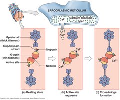
Steps in Muscle Contraction
Ca2+ influx exposes actin active sites.
Myosin cross bridge forms with actin.
Power stroke moves thin filament (uses ATP).
ATP binds to myosin, breaking cross bridge.
ATP hydrolysis re-energizes myosin.
Ca2+ returns to SR, actin sites covered, muscle relaxes.
Muscle Relaxation
AChE breaks down ACh at NMJ.
ATP breaks myosin-actin cross bridge.
Ca2+ returns to SR.
Muscle Tension and Contraction Types
All-or-None Principle
Muscle fiber contracts fully or not at all.
Tension depends on number of cross bridges, sarcomere length, stimulation frequency, and number of fibers stimulated.
Length-Tension Relationship
Optimal sarcomere length produces maximum tension.
Too much or too little overlap reduces tension.
Muscle Twitch and Contraction Types
Muscle twitch: Single contraction-relaxation cycle (latent, contraction, relaxation phases).
Treppe: Successive contractions increase tension.
Wave summation: Rapid stimuli add tension.
Incomplete tetanus: Rapid cycles, almost peak contraction.
Complete tetanus: No relaxation, maximum tension.
Motor Units
All muscle fibers controlled by one motor neuron.
Small motor units: fine control (e.g., eye muscles).
Large motor units: powerful movements (e.g., leg muscles).
Types of Contractions
Isotonic: Tension constant, length changes.
Concentric: Muscle shortens.
Eccentric: Muscle lengthens.
Isometric: Length unchanged, tension maintained.
Muscle Energetics
ATP Sources
Creatine phosphate: Transfers phosphate to ADP via creatine kinase.
Glycolysis: Anaerobic, produces 2 ATP/glucose, lactic acid as byproduct.
Aerobic metabolism: Uses O2 and glucose, produces most ATP.
Myoglobin: Stores O2 in muscle.
Oxygen Debt and Fatigue
Oxygen debt: Increased O2 needed post-exercise (EPOC).
Muscle fatigue: Depletion of reserves, damage, pH decline, pain.
Heat Production
Muscle contraction generates heat (e.g., shivering).
Types of Skeletal Muscle Fibers
Fast, Slow, and Intermediate Fibers
Fast twitch: Powerful, fatigue quickly, anaerobic.
Slow twitch: Endurance, fatigue-resistant, aerobic, high myoglobin.
Intermediate: Properties between fast and slow, moderate fatigue resistance.
Muscle Tone and Adaptation
Muscle Tone
Hypertrophy: Increase in cell diameter, muscle mass.
Atrophy: Decrease in size and strength due to inactivity.
Cardiac and Smooth Muscle
Cardiac Muscle
Involuntary, striated, branched cells, intercalated disks.
Uses aerobic metabolism, contracts without neural stimulus.
Smooth Muscle
Involuntary, non-striated, spindle-shaped cells.
Lacks T-tubules and sarcomeres.
Muscle Fascicle Arrangements
Types of Fascicle Arrangements
Parallel: Fascicles run parallel (e.g., biceps brachii).
Convergent: Broad area converges on tendon (e.g., pectoralis major).
Pennate: Fascicles at angle to tendon (unipennate, bipennate, multipennate).
Circular: Fascicles form rings (sphincters).
Levers in Muscle Action
Classes of Levers
First class: Fulcrum between load and force (e.g., pry-bar).
Second class: Load between force and fulcrum (e.g., wheelbarrow).
Third class: Force between load and fulcrum (e.g., catapult).
Muscle Diseases (For Reference)
Muscular Dystrophy
Genetic disorder causing progressive muscle weakness.
No cure; management with medication and therapy.
Myasthenia Gravis
Autoimmune disease affecting NMJ, causing muscle weakness.
No cure; treatment manages symptoms.
Additional info: Expanded explanations, definitions, and examples were added for clarity and completeness. The included image directly illustrates the role of calcium ions in muscle contraction, showing the sarcoplasmic reticulum, exposure of actin sites, and cross-bridge formation, which is central to the sliding filament theory and excitation-contraction coupling.
