 Indietro
Chapter 10: The Muscular System – Structure, Function, and Major Muscles
Guida di studio - Note intelligenti
 Appunti personalizzati basati sui tuoi materiali, ampliati con definizioni chiave, esempi e contesto.
Muscle Actions and Interactions
Functional Groups of Muscles
Muscles work together in groups to produce, control, and stabilize movements. Understanding these groups is essential for analyzing muscle function.
Prime mover (agonist): The muscle primarily responsible for generating a specific movement. Example: Biceps brachii during elbow flexion.
Antagonist: Opposes or reverses the action of the prime mover. Example: Triceps brachii during elbow flexion.
Synergist: Assists the prime mover by adding force or reducing unnecessary movements.
Fixator: A type of synergist that stabilizes the origin of the prime mover, providing a stable base.
Example: During flexion of the forearm, the biceps brachii is the prime mover, the triceps brachii is the antagonist, and muscles like the brachialis act as synergists.
Muscle Position Relative to Joints
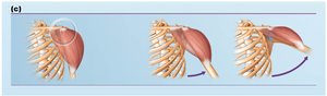
The action of a muscle can often be inferred from its position relative to the joint it crosses:
Anterior side: Produces flexion (except at knee and ankle).
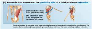
Posterior side: Produces extension (except at knee and ankle).
Lateral side: Produces abduction.
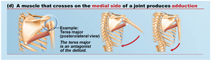
Medial side: Produces adduction.
Example: The latissimus dorsi crosses the posterior side of the shoulder joint and produces extension. The deltoid crosses the lateral side and produces abduction. The teres major crosses the medial side and produces adduction.
Naming Skeletal Muscles
Criteria for Muscle Naming
Skeletal muscles are named based on several criteria, which often provide clues to their location, shape, and function:
Location: Indicates the bone or region associated (e.g., temporalis over temporal bone).
Shape: Describes the muscle's form (e.g., deltoid = triangle, trapezius = trapezoid).
Size: Terms like maximus (largest), minimus (smallest), longus (long), brevis (short).
Direction of fibers: Rectus (straight), transversus (right angles), oblique (angles).
Number of origins: Biceps (two), triceps (three).
Location of attachments: Origin and insertion points (e.g., sternocleidomastoid).
Action: Flexor, extensor, adductor, abductor.
Example: Extensor carpi radialis longus – extends the wrist (carpi), is associated with the radius, and is long.
Fascicle Arrangements
Types of Fascicle Arrangements
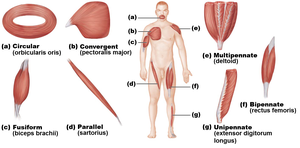
Fascicles are bundles of muscle fibers, and their arrangement determines muscle shape and function. The main patterns are:
Circular: Fascicles arranged in concentric rings (e.g., orbicularis oris).
Convergent: Broad origin, fascicles converge to a single tendon (e.g., pectoralis major).
Parallel: Fascicles run parallel to the long axis (e.g., sartorius).
Fusiform: Spindle-shaped with parallel fibers (e.g., biceps brachii).
Pennate: Short fascicles attach obliquely to a central tendon. Types include unipennate, bipennate, and multipennate (e.g., rectus femoris, deltoid).
Functional Implications of Fascicle Arrangement
The arrangement affects both the range of motion and power:
Range of motion: Muscles with long, parallel fibers shorten more, allowing greater movement.
Power: Muscles with more fibers (e.g., bipennate, multipennate) are more powerful but shorten less.
Example: The deltoid (multipennate) is powerful, while the sartorius (parallel) allows greater movement.
Major Skeletal Muscles of the Body
Overview and Learning Tips
There are over 600 skeletal muscles, grouped by function and location. Muscle tables typically include description, origin and insertion, actions, and innervation.
Relate muscle names to their location and function.
Identify muscles on anatomical figures.
Act out movements to understand muscle actions.
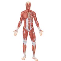
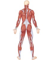
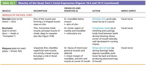
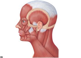
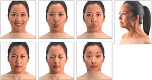
Muscles of the Head: Facial Expression
Facial Expression Muscles
Muscles of facial expression insert into the skin, not bone, and are crucial for nonverbal communication. All are innervated by cranial nerve VII (facial nerve).
Muscles of the scalp: e.g., epicranius
Muscles of the face: e.g., orbicularis oculi, zygomaticus, orbicularis oris
Muscles of Mastication and Tongue Movement
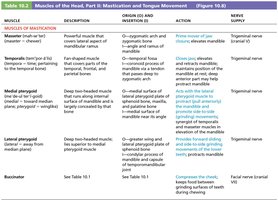
Mastication Muscles
Four pairs of muscles, all innervated by cranial nerve V, control jaw movements:
Prime movers: Temporalis and masseter (jaw closure)
Grinding movements: Pterygoids
Chewing role: Buccinator
Tongue Movement Muscles
Three extrinsic muscles anchor and move the tongue: genioglossus, hyoglossus, styloglossus.
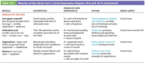
Summary Table: Muscles of the Head, Part I: Facial Expression
Muscle | Description | Origin (O) and Insertion (I) | Action | Nerve Supply |
|---|---|---|---|---|
Corrugator supercilii | Small muscle; activity associated with frowning | O: arch of frontal bone above nasal bone I: skin of eyebrow | Draws eyebrows medially and inferiorly | Facial nerve |
Orbicularis oculi | Thin, tripartite muscle of eyelids; surrounds rim of orbit | O: frontal and maxillary bones and ligaments around orbit I: tissue of eyelid | Closes eye; produces blinking and squinting | Facial nerve |
Zygomaticus (major and minor) | Muscle pair extending diagonally from cheekbone to corner of mouth | O: zygomatic bone I: skin and muscle at corner of mouth | Raises lateral corners of mouth (smiling) | Facial nerve |
Risorius | Slender muscle inferior and lateral to zygomaticus | O: lateral fascia associated with masseter muscle I: skin at angle of mouth | Draws corner of lip laterally | Facial nerve |
Summary Table: Muscles of Mastication
Muscle | Description | Origin (O) and Insertion (I) | Action | Nerve Supply |
|---|---|---|---|---|
Masseter | Powerful muscle; covers lateral aspect of mandibular ramus | O: zygomatic arch I: angle and ramus of mandible | Prime mover of jaw closure | Trigeminal nerve |
Temporalis | Fan-shaped muscle; covers parts of temporal, frontal, and parietal bones | O: temporal fossa I: coronoid process of mandible | Closes jaw; elevates and retracts mandible | Trigeminal nerve |
Medial pterygoid | Deep two-headed muscle; runs along internal surface of mandible | O: medial surface of lateral pterygoid plate I: medial surface of mandible | Promotes side-to-side grinding movements | Trigeminal nerve |
Lateral pterygoid | Deep two-headed muscle; lies superior to medial pterygoid | O: greater wing and lateral pterygoid plate I: condylar process of mandible | Provides forward sliding and side-to-side grinding movements | Trigeminal nerve |
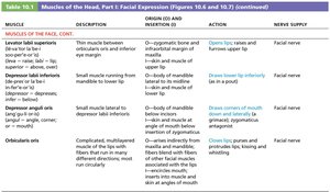
Buccinator | Deep muscle; forms lateral wall of oral cavity | See Table 10.1 I: blends with fibers of orbicularis oris | Compresses cheek; keeps food between grinding surfaces of teeth during chewing | Facial nerve |
