 Indietro
Chapter 11: Fundamentals of the Nervous System and Nervous Tissue – Study Notes
Guida di studio - Note intelligenti
 Appunti personalizzati basati sui tuoi materiali, ampliati con definizioni chiave, esempi e contesto.
Fundamentals of the Nervous System
Functions of the Nervous System
The nervous system is the master controlling and communicating system of the body. It utilizes electrical and chemical signals to coordinate rapid and specific responses, often resulting in immediate actions.
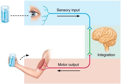
Sensory Input: Information is gathered by sensory receptors about internal and external changes.
Integration: The processing and interpretation of sensory input occurs in the central nervous system.
Motor Output: Activation of effector organs (muscles and glands) produces a response.
Example: Seeing a glass of water (sensory input), the brain processes this information (integration), and the arm moves to pick up the glass (motor output).
Organization of the Nervous System
Central and Peripheral Nervous Systems
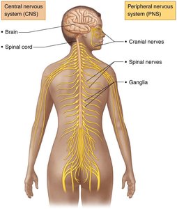
The nervous system is divided into two principal parts: the Central Nervous System (CNS) and the Peripheral Nervous System (PNS).
Central Nervous System (CNS): Consists of the brain and spinal cord, located in the dorsal body cavity. It serves as the integration and control center, interpreting sensory input and dictating motor output.
Peripheral Nervous System (PNS): Comprises nerves that extend from the brain and spinal cord. It includes spinal nerves (to and from the spinal cord) and cranial nerves (to and from the brain).
Functional Divisions of the PNS
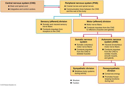
The PNS is further divided into sensory (afferent) and motor (efferent) divisions, each with specialized functions.
Sensory (Afferent) Division: Somatic sensory fibers convey impulses from skin, skeletal muscles, and joints to the CNS. Visceral sensory fibers convey impulses from visceral organs to the CNS.
Motor (Efferent) Division: Transmits impulses from the CNS to effector organs (muscles and glands). It is subdivided into:
Somatic Nervous System: Conducts impulses from CNS to skeletal muscle; voluntary control.
Autonomic Nervous System (ANS): Regulates smooth muscle, cardiac muscle, and glands; involuntary control. The ANS is further divided into:
Sympathetic Division: Mobilizes body systems during activity.
Parasympathetic Division: Conserves energy and promotes housekeeping functions during rest.
Histology of Nervous Tissue
Principal Cell Types
Nervous tissue consists of two main cell types: neuroglia (glial cells) and neurons (nerve cells).
Neuroglia: Small cells that surround and wrap delicate neurons, providing support and protection.
Neurons: Excitable cells that transmit electrical signals.
Neuroglia of the CNS
Four main types of neuroglia support CNS neurons:
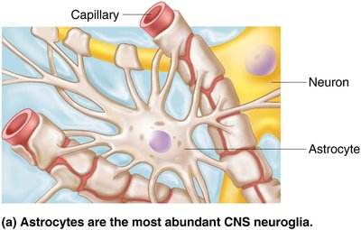
Astrocytes: Most abundant, versatile, and highly branched glial cells. They support and brace neurons, facilitate exchanges between capillaries and neurons, guide migration of young neurons, control the chemical environment, respond to nerve impulses, and participate in information processing.
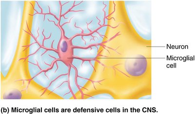
Microglial Cells: Small, ovoid cells with thorny processes. They monitor neurons, migrate toward injured neurons, and can transform to phagocytize microorganisms and neuronal debris.
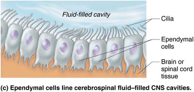
Ependymal Cells: Range in shape from squamous to columnar and may be ciliated. Their cilia circulate cerebrospinal fluid (CSF), and they line the central cavities of the brain and spinal column, forming a permeable barrier between CSF and tissue fluid.
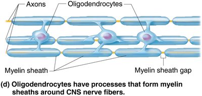
Oligodendrocytes: Branched cells whose processes wrap CNS nerve fibers, forming insulating myelin sheaths in thicker nerve fibers.
Neuroglia of the PNS
Two major neuroglia are found in the PNS:
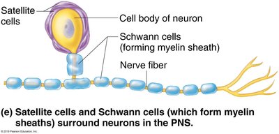
Satellite Cells: Surround neuron cell bodies in the PNS and function similarly to astrocytes in the CNS.
Schwann Cells (Neurolemmocytes): Surround all peripheral nerve fibers and form myelin sheaths in thicker nerve fibers. They are vital to the regeneration of damaged peripheral nerve fibers.
Neurons: Structure and Function
Neuron Cell Body
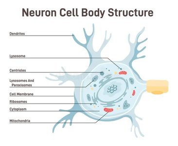
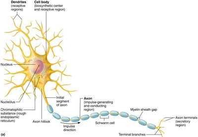
The neuron cell body, also called the perikaryon or soma, is the biosynthetic center of the neuron. It synthesizes proteins, membranes, and chemicals, and contains a spherical nucleus with a nucleolus. Most neuron cell bodies are located in the CNS (nuclei), while clusters in the PNS are called ganglia.
Neuron Processes
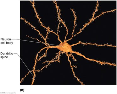
Neurons have armlike processes that extend from the cell body. The CNS contains both neuron cell bodies and their processes, while the PNS contains chiefly neuron processes. Bundles of neuron processes in the CNS are called tracts, and in the PNS are called nerves. There are two types of processes: dendrites and axons.
Dendrites: Short, tapering, diffusely branched processes that serve as the receptive (input) region of the neuron. They convey incoming messages toward the cell body as graded potentials.
Axon: Each neuron has one axon that starts at the axon hillock. Axons can be very long and are called nerve fibers. They branch profusely at their end, forming terminal branches and axon terminals (terminal boutons).
Functional Characteristics: The axon is the conducting region of the neuron, generating and transmitting nerve impulses along the axolemma to the axon terminal. Axons rely on cell bodies to renew proteins and membranes and decay quickly if cut or damaged.
Internal Transport: Axons have efficient internal transport mechanisms, moving molecules and organelles via motor proteins and cytoskeletal elements. Movement occurs in both directions: anterograde (away from cell body) and retrograde (toward cell body).
Myelin Sheath
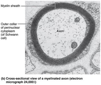
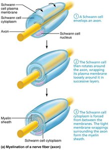
The myelin sheath is composed of myelin, a whitish, protein-lipid substance. Its function is to protect and electrically insulate the axon, increasing the speed of nerve impulse transmission. Myelinated fibers have segmented sheaths, while nonmyelinated fibers conduct impulses more slowly.
Myelination in the PNS: Formed by Schwann cells, which wrap around the axon in a jelly roll fashion. One cell forms one segment of the myelin sheath. The outer collar of perinuclear cytoplasm contains the nucleus and most of the cytoplasm.
Myelin Sheath Gaps: Gaps between adjacent Schwann cells, formerly called nodes of Ranvier, are sites where axon collaterals can emerge.
Nonmyelinated Fibers: Thin fibers not wrapped in myelin; surrounded by Schwann cells but no coiling.
Myelin Sheaths in the CNS: Formed by processes of oligodendrocytes. Each cell can wrap up to 60 axons at once. White matter consists of myelinated fibers, while gray matter is mostly neuron cell bodies and nonmyelinated fibers.
Classification of Neurons
Functional Classification
Neurons are classified by the direction in which nerve impulses travel relative to the CNS:
Sensory (Afferent) Neurons: Transmit impulses from sensory receptors toward the CNS. Almost all are unipolar, and their cell bodies are located in ganglia in the PNS.
Motor (Efferent) Neurons: Carry impulses from the CNS to effectors. They are multipolar, and most cell bodies are located in the CNS (except some autonomic neurons).
Interneurons (Association Neurons): Lie between motor and sensory neurons, shuttling signals through CNS pathways. Most are entirely within the CNS, and they constitute 99% of the body's neurons.
Type | Direction of Impulse | Location of Cell Body | Structure |
|---|---|---|---|
Sensory (Afferent) | Toward CNS | PNS (Ganglia) | Unipolar |
Motor (Efferent) | Away from CNS | CNS (except some autonomic) | Multipolar |
Interneurons | Within CNS | CNS | Multipolar |
Additional info: The functional classification of neurons is essential for understanding how information is processed and transmitted throughout the nervous system, enabling complex behaviors and responses.
