 Indietro
Chapter 11: Fundamentals of the Nervous System and Nervous Tissue – Study Guide
Guida di studio - Note intelligenti
 Appunti personalizzati basati sui tuoi materiali, ampliati con definizioni chiave, esempi e contesto.
Nervous System Overview
Functions of the Nervous System
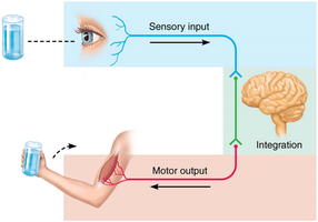
The nervous system is responsible for detecting stimuli, processing information, and initiating responses. It performs three major functions:
Sensory Input: Gathering information from sensory receptors about internal and external environments.
Integration: Processing and interpreting sensory information to determine an appropriate response.
Motor Output: Activating effector organs (muscles and glands) to produce a response.
Example: Touching a hot stove: Sensory receptors detect heat, CNS interprets the stimulus, and muscles pull the hand away.
Organization of the Nervous System
Major Divisions
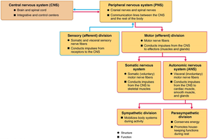
The nervous system is divided into two main parts:
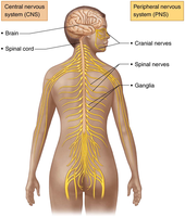
Central Nervous System (CNS): Consists of the brain and spinal cord. It is the integration and control center, interpreting sensory input and dictating motor output.
Peripheral Nervous System (PNS): Includes all nervous tissue outside the CNS, mainly nerves that extend from the brain and spinal cord. Spinal nerves carry impulses to and from the spinal cord; cranial nerves carry impulses to and from the brain.
Functional Divisions of the PNS
The PNS is further divided based on function:
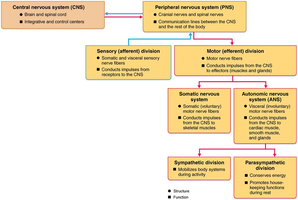
Sensory (Afferent) Division: Carries information to the CNS. Includes somatic sensory fibers (from skin, muscles, joints) and visceral sensory fibers (from organs).
Motor (Efferent) Division: Carries commands from the CNS to effector organs. Includes the somatic nervous system (voluntary control of skeletal muscles) and the autonomic nervous system (involuntary control of smooth muscle, cardiac muscle, and glands).
Central Nervous System (CNS)
CNS Functions and Components
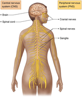
The CNS receives sensory information, processes it, makes decisions, and directs motor responses. Its main components are:
Brain: The primary organ for integration and processing.
Spinal Cord: Conducts information between the brain and the rest of the body.
Peripheral Nervous System (PNS)
PNS Components and Function
The PNS acts as the communication highway between the CNS and the rest of the body. Its components include:
Cranial Nerves: Carry information to and from the brain.
Spinal Nerves: Carry information to and from the spinal cord.
Ganglia: Clusters of neuron cell bodies in the PNS.
Functional Divisions of the PNS
Sensory and Motor Divisions
The PNS is divided into sensory and motor divisions:
Sensory Division (Afferent): Arrives at the CNS with information from somatic and visceral sources.
Motor Division (Efferent): Exits the CNS with commands to muscles and glands. Includes:
Somatic Nervous System: Voluntary control of skeletal muscles.
Autonomic Nervous System: Involuntary control of smooth muscle, cardiac muscle, and glands. Divided into sympathetic (fight or flight) and parasympathetic (rest and digest) divisions.
Nervous Tissue
Cell Types
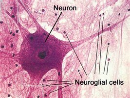
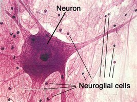
Nervous tissue consists of two major cell types:
Neurons: Excitable cells that transmit electrical signals. Most are formed before birth and last a lifetime; mature neurons rarely divide.
Neuroglia (Glial Cells): Support, protect, insulate, and nourish neurons. Neuroglia outnumber neurons.
Neuroglia Overview
Neuroglia in CNS and PNS
Neuroglia are essential for neuron function and maintenance. In the CNS, four main types support neurons:
Astrocytes: Most abundant; support neurons, regulate chemical environment, guide neuron migration, and participate in information processing.
Microglia: Monitor neurons, migrate toward injury, and phagocytize debris.
Ependymal Cells: Line brain and spinal cord cavities; circulate cerebrospinal fluid (CSF).
Oligodendrocytes: Form myelin sheaths in CNS.
In the PNS:
Satellite Cells: Surround neuron cell bodies in ganglia; similar to astrocytes.
Schwann Cells: Form myelin sheaths around peripheral nerve fibers; vital for regeneration.
Neuron Structure and Function
Neuron Overview
Neurons are the structural units of the nervous system. They are large, highly specialized cells that conduct impulses. Key characteristics:
Extreme Longevity: Last a person’s lifetime.
Amitotic: With few exceptions, do not divide after maturity.
High Metabolic Rate: Require continuous supply of oxygen and glucose.
Structure: All have a cell body (soma) and one or more processes (dendrites and axons).
Neuron Cell Body
The cell body (soma) is the biosynthetic center of the neuron, containing mitochondria, rough ER (Nissl bodies), and a nucleus. It synthesizes proteins, membranes, and chemicals necessary for neuron function and repair.
Nuclei vs Ganglia
Nuclei: Clusters of neuron cell bodies in the CNS.
Ganglia: Clusters of neuron cell bodies in the PNS.
Neuron Processes
Neurons have two types of processes:
Dendrites: Short, branched processes that receive input and convey messages toward the cell body as graded potentials.
Axon: A single, long process that conducts impulses away from the cell body. Axons can branch and end in axon terminals, which release neurotransmitters.
Tracts vs Nerves
Tracts: Bundles of axons in the CNS.
Nerves: Bundles of axons in the PNS.
Myelin Sheath
Structure and Function
The myelin sheath is a segmented, protein-lipid covering that insulates axons and increases the speed of nerve impulse transmission.
Myelinated Fibers: Have a myelin sheath; conduct impulses rapidly.
Nonmyelinated Fibers: Lack a sheath; conduct impulses more slowly.
Myelination in the PNS
Schwann cells wrap around axons in a jelly roll fashion, forming the myelin sheath. Gaps between Schwann cells are called Nodes of Ranvier, which allow action potentials to jump rapidly.
Myelination in the CNS
Oligodendrocytes form myelin sheaths in the CNS. White matter consists of myelinated fibers (tracts), while gray matter contains cell bodies, dendrites, and unmyelinated fibers.
Types of Neurons – Structural Classification
Multipolar, Bipolar, and Unipolar Neurons
Multipolar: Three or more processes (one axon, multiple dendrites); most common in CNS.
Bipolar: Two processes (one axon, one dendrite); rare, found in retina and olfactory mucosa.
Unipolar: One T-like process; found mainly in PNS ganglia, associated with sensory reception.
Types of Neurons – Functional Classification
Sensory, Motor, and Interneurons
Sensory (Afferent): Transmit impulses from sensory receptors toward CNS; mostly unipolar, cell bodies in PNS ganglia.
Motor (Efferent): Carry impulses away from CNS to effectors; exiting the CNS.
Interneurons: Association neurons; shuttle signals within CNS, most abundant.
Resting Membrane Potential
Ion Concentration and Permeability
Neurons have a resting membrane potential, typically around −70 mV. The inside of the cell is negative relative to the outside. This potential is generated by:
Differences in ionic composition: ECF has high Na+, low K+; ICF has high K+, low Na+, and negatively charged proteins.
Differences in membrane permeability: Membrane is more permeable to K+ than Na+.
At rest, K+ movement dominates, making the inside more negative.
Membrane Ion Channels
Chemically gated (ligand-gated): Open with binding of a specific chemical.
Voltage-gated: Open and close in response to changes in membrane potential.
Mechanically gated: Open and close in response to physical deformation.
Changes in Membrane Potential
Depolarization and Hyperpolarization
Depolarization: Membrane potential decreases, inside becomes less negative; increases probability of producing an impulse.
Hyperpolarization: Membrane potential increases, inside becomes more negative; decreases probability of producing an impulse.
Action Potentials
Generation and Propagation
Action potentials are brief, long-distance signals within neurons. They occur only in excitable membranes and involve a rapid reversal of membrane potential (~100 mV).
Resting State: All gated channels closed; membrane potential maintained.
Depolarization: Na+ channels open, Na+ enters, membrane potential rises to +30 mV.
Repolarization: Na+ channels close, K+ channels open, K+ exits, membrane potential returns toward resting.
Hyperpolarization: K+ channels stay open briefly, membrane potential dips below resting before returning to normal.
Action potentials are self-propagating and do not decay over distance. The refractory period ensures unidirectional flow.
Threshold & All-or-None Phenomenon
Threshold: Depolarization must reach a certain voltage to trigger an action potential.
All-or-None: An action potential either happens completely or not at all.
Propagation of Action Potential
Once initiated, the action potential travels down the axon toward the terminals, triggering voltage-gated channels in adjacent regions.
Coding for Stimulus Intensity
Stimulus intensity is coded by the frequency of action potentials, not their size.
Conduction Velocity
Saltatory Conduction: Occurs in myelinated axons; action potentials jump from node to node, increasing speed.
Synapses
Transmission Between Neurons
Synapses are junctions that mediate information transfer between neurons or from neurons to effector cells. Most are chemical synapses, involving neurotransmitter release and reception.
Presynaptic Neuron: Sends information.
Postsynaptic Neuron: Receives information.
Chemical Synaptic Transmission
Action potential arrives at axon terminal.
Voltage-gated Ca2+ channels open; Ca2+ enters.
Ca2+ triggers synaptic vesicles to release neurotransmitter by exocytosis.
Neurotransmitter binds to receptors on postsynaptic membrane.
Ion channels open, producing graded potentials (excitatory or inhibitory).
Neurotransmitter effects end by reuptake, enzymatic degradation, or diffusion.
Student Questions & Additional Info
Heart vs Skeletal Muscle Action Potentials
Skeletal Muscle: Requires nervous system stimulation; action potential begins in a somatic motor neuron.
Cardiac Muscle: Generates its own action potentials via pacemaker cells; autonomic nerves modulate rate but do not initiate each beat.
Astroblast Cell
An astroblast is an immature astrocyte precursor, actively dividing and differentiating during CNS development.
In-Class Activity
Draw a neuron and show how a message is received and passed to the next cell, including dendrites, cell body, axon, action potential graph, axon terminal, synaptic cleft, and next cell.
