 Indietro
Chapter 11: The Cardiovascular System – Structure, Function, and Physiology
Guida di studio - Note intelligenti
 Appunti personalizzati basati sui tuoi materiali, ampliati con definizioni chiave, esempi e contesto.
The Cardiovascular System: Overview
Major Functions and Importance
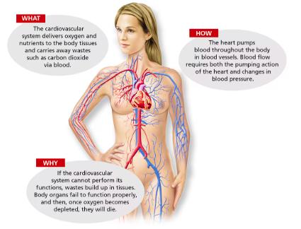
The cardiovascular system is essential for transporting oxygen, nutrients, hormones, and waste products throughout the body. It maintains homeostasis by ensuring that all body tissues receive the substances they need and that metabolic wastes are efficiently removed.
Transportation: Blood serves as the transport medium, carrying oxygen, nutrients, and hormones to cells and removing carbon dioxide and other wastes.
Force Generation: The heart acts as a muscular pump, generating the pressure required to move blood through the vessels.
Homeostasis: Proper cardiovascular function is vital for tissue survival; failure leads to waste accumulation and organ dysfunction.
Anatomy of the Heart
Size, Location, and Orientation
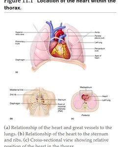
The heart is located in the mediastinum, the central compartment of the thoracic cavity, flanked by the lungs. Its apex points toward the left hip and rests on the diaphragm, while its base lies beneath the second rib, directed toward the right shoulder.
Apex: Directed toward the left hip, at the level of the 5th intercostal space.
Base: Oriented toward the right shoulder, beneath the 2nd rib.
Coverings and Walls of the Heart
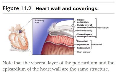
The heart is enclosed by the pericardium, a triple-layered sac that protects and anchors the heart. The outer fibrous pericardium provides protection and attachment, while the inner serous pericardium (parietal and visceral layers) produces lubricating fluid to reduce friction during heartbeats.
Epicardium (Visceral Pericardium): Outermost layer of the heart wall and innermost layer of the pericardium.
Myocardium: Thick, contractile middle layer composed of cardiac muscle cells interconnected by intercalated discs.
Endocardium: Thin, smooth inner lining continuous with blood vessel endothelium.
Gross Anatomy of the Heart
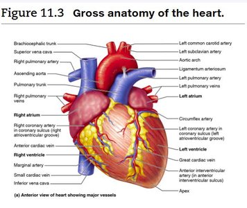
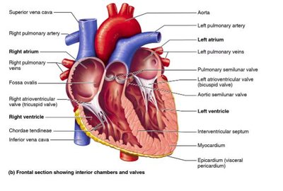
The heart consists of four chambers: two atria (receiving chambers) and two ventricles (pumping chambers). The right side pumps blood to the lungs (pulmonary circuit), while the left side pumps blood to the body (systemic circuit).
Right Atrium & Ventricle: Receive deoxygenated blood and pump it to the lungs.
Left Atrium & Ventricle: Receive oxygenated blood from the lungs and pump it to the body.
Septum: Divides the heart longitudinally into right and left sides (interatrial and interventricular septa).
Circulatory Pathways: Pulmonary and Systemic Circuits
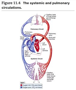
The heart functions as a double pump. The right side (pulmonary circuit) sends blood to the lungs for oxygenation, while the left side (systemic circuit) delivers oxygen-rich blood to the body tissues.
Pulmonary Circuit: Right ventricle → pulmonary trunk → pulmonary arteries → lungs → pulmonary veins → left atrium.
Systemic Circuit: Left ventricle → aorta → systemic arteries → body tissues → systemic veins → right atrium.
Anatomical Differences in Ventricles
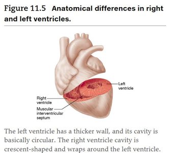
The left ventricle has a thicker wall and a more circular cavity compared to the right ventricle, which is crescent-shaped and wraps around the left ventricle. This structural difference allows the left ventricle to generate higher pressure for systemic circulation.
Heart Valves
Types and Functions
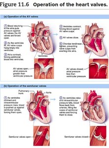
The heart contains four valves that ensure unidirectional blood flow:
Atrioventricular (AV) Valves: Located between atria and ventricles (tricuspid on right, bicuspid/mitral on left). Prevent backflow into atria during ventricular contraction.
Semilunar Valves: Located at the bases of the large arteries leaving the ventricles (pulmonary and aortic semilunar valves). Prevent backflow into ventricles after contraction.
Chordae Tendineae: Tendinous cords anchoring AV valve cusps to ventricular walls, preventing prolapse.
Valve Disorders and Replacement
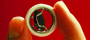
Valve dysfunction can lead to backflow (incompetence) or narrowing (stenosis), increasing cardiac workload and potentially leading to heart failure. Severely damaged valves may be replaced with synthetic or biological prostheses.
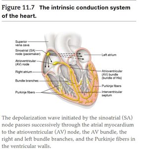
Intrinsic Conduction System of the Heart
Components and Function
The intrinsic conduction system (nodal system) coordinates the heart's rhythmic contractions. It consists of specialized cardiac tissue that generates and conducts electrical impulses.
Sinoatrial (SA) Node: Pacemaker of the heart, initiates each heartbeat.
Atrioventricular (AV) Node: Delays impulse, allowing atrial contraction before ventricular contraction.
AV Bundle (Bundle of His), Bundle Branches, Purkinje Fibers: Distribute impulse through ventricles, ensuring coordinated contraction.
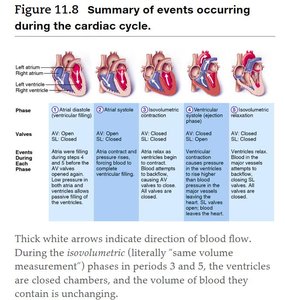
Cardiac Cycle and Heart Sounds
Phases of the Cardiac Cycle
The cardiac cycle consists of all events in one heartbeat, including atrial and ventricular contraction (systole) and relaxation (diastole). The sequence ensures efficient blood flow through the heart and into the circulation.
Atrial Diastole (Ventricular Filling): AV valves open, blood flows into ventricles.
Atrial Systole: Atria contract, completing ventricular filling.
Isovolumetric Contraction: Ventricles contract with all valves closed, building pressure.
Ventricular Systole (Ejection Phase): Semilunar valves open, blood ejected into arteries.
Isovolumetric Relaxation: Ventricles relax, semilunar valves close, AV valves reopen as pressure drops.
Heart Sounds
Two main heart sounds are heard during each cardiac cycle:
"Lub": Closure of AV valves at the start of ventricular systole.
"Dup": Closure of semilunar valves at the end of ventricular systole.
Abnormal sounds (murmurs) may indicate valve problems.
Cardiac Output and Its Regulation
Definition and Calculation
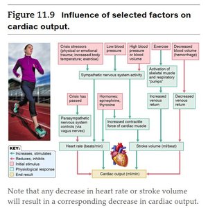
Cardiac output (CO) is the volume of blood pumped by each ventricle per minute. It is calculated as:
HR: Heart rate (beats per minute)
SV: Stroke volume (ml per beat)
Normal adult CO ≈ 5250 ml/min (75 bpm × 70 ml/beat).
Factors Influencing Cardiac Output
Preload: Degree of stretch of cardiac muscle before contraction (Frank-Starling law).
Contractility: Strength of contraction, influenced by calcium, sympathetic stimulation, and hormones.
Afterload: Pressure the ventricles must overcome to eject blood.
Blood Vessels: Structure and Function
Types of Blood Vessels
Blood vessels form a closed circuit for blood flow. The main types are:
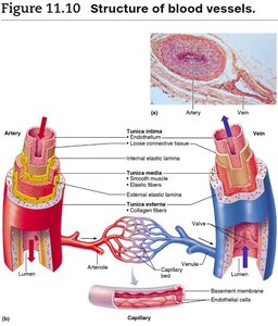
Arteries: Carry blood away from the heart; thick-walled, high pressure.
Veins: Return blood to the heart; thinner walls, larger lumens, often contain valves.
Capillaries: Microscopic vessels where exchange of gases, nutrients, and wastes occurs; walls are one cell thick.
Venous Return Mechanisms
Venous return is aided by:
Valves: Prevent backflow in veins.
Muscular Pump: Skeletal muscle contractions "milk" blood toward the heart.
Respiratory Pump: Pressure changes during breathing help draw blood toward the heart.
Summary Table: Key Structural Differences in Blood Vessels
Feature | Arteries | Veins | Capillaries |
|---|---|---|---|
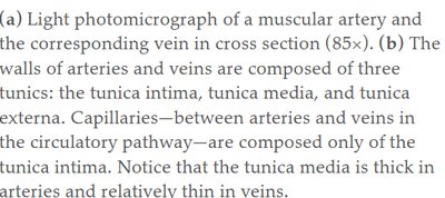
Wall Thickness | Thick (especially tunica media) | Thin | One cell layer (endothelium) |
Lumen Size | Small | Large | Very small |
Valves | Absent | Present (in limbs) | Absent |
Function | Carry blood away from heart | Return blood to heart | Exchange of substances |
Additional info:
Heart murmurs are often benign in children but may indicate pathology in adults.
Congestive heart failure can result from chronic hypertension, coronary artery disease, or myocardial infarction.
Regular aerobic exercise strengthens the heart and improves vascular health.
