 Indietro
Chapter 12: Muscle Physiology (Skeletal Muscle Focus)
Guida di studio - Note intelligenti
 Appunti personalizzati basati sui tuoi materiali, ampliati con definizioni chiave, esempi e contesto.
Muscle Physiology: Structure and Function
Skeletal Muscle Structure
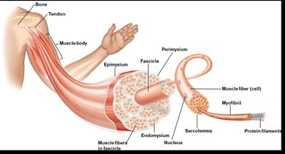
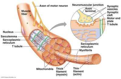
Skeletal muscle is composed of bundles of muscle fibers, each surrounded by connective tissue and organized for efficient contraction. The sarcolemma is the plasma membrane of the muscle cell, and the sarcoplasm is its cytoplasm. Myofibrils are the contractile organelles within muscle fibers, responsible for generating force.
Epimysium, Perimysium, Endomysium: Layers of connective tissue that surround the muscle body, fascicles, and individual fibers, respectively.
Myofibril: Runs the length of the myocyte and contains the contractile proteins.
Sarcolemma: Muscle cell membrane.
Sarcoplasm: Muscle cell cytoplasm.
Muscle Fiber Organelles
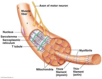
Muscle fibers contain specialized organelles for contraction and energy production. The sarcoplasmic reticulum (SR) stores calcium ions, which are essential for muscle contraction. Transverse tubules (T-tubules) are invaginations of the sarcolemma that help transmit action potentials deep into the muscle fiber.
Sarcoplasmic Reticulum (SR): Surrounds myofibrils and stores Ca++.
Terminal Cisternae: Enlarged regions of the SR that contact T-tubules.
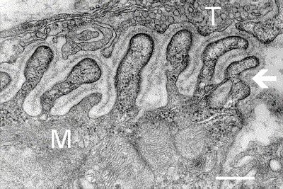
T-tubules: Invaginations of the sarcolemma for action potential transmission.
The Sarcomere: Functional Unit of Muscle
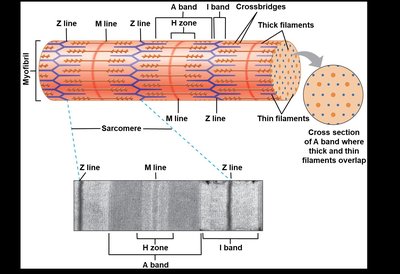
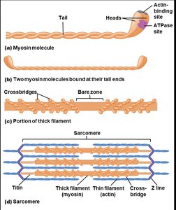
The sarcomere is the basic contractile unit of muscle, defined by the arrangement of actin (thin) and myosin (thick) filaments. This organization gives skeletal muscle its striated appearance.
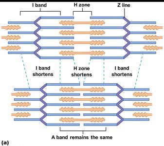
Z line: Defines the boundaries of a sarcomere.
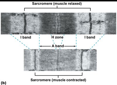
A band: Region containing thick filaments.
I band: Region containing only thin filaments.
H zone: Region containing only thick filaments.
Crossbridges: Connections formed between actin and myosin during contraction.
Muscle Contraction Mechanisms
Contractile and Regulatory Proteins
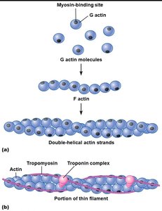
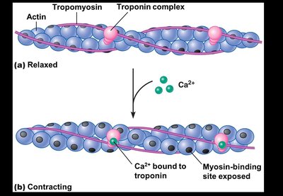
Muscle contraction is driven by the interaction of contractile proteins (actin and myosin) and regulated by troponin and tropomyosin. Actin forms the thin filament, while myosin forms the thick filament.
Actin: G actin monomers polymerize to form F actin, which makes up the thin filament.
Myosin: Each molecule has a head (with ATPase and actin-binding sites) and a tail.
Tropomyosin: Covers myosin-binding sites on actin in relaxed muscle.
Troponin: Binds Ca++ and moves tropomyosin to expose binding sites.
Sliding Filament Mechanism
Muscle contraction occurs when myosin heads bind to actin and pull the thin filaments toward the center of the sarcomere, shortening the muscle.
During contraction: I band and H zone shorten, A band remains constant.
Crossbridge cycling: Myosin heads repeatedly attach, pull, and detach from actin.
The Crossbridge Cycle
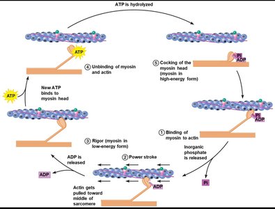
The crossbridge cycle describes the sequence of events that occur during muscle contraction, powered by ATP hydrolysis.
Binding of myosin to actin
Power stroke: Myosin head pivots, pulling actin.
Rigor: Myosin remains bound to actin (no ATP).
Unbinding: ATP binds to myosin, causing release from actin.
Cocking: ATP hydrolysis re-energizes myosin head.
Excitation-Contraction Coupling
Neuromuscular Junction (NMJ)
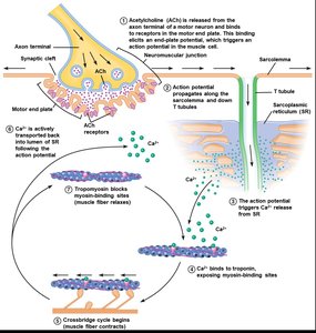
The NMJ is the synapse between a somatic motor neuron and a muscle fiber. Acetylcholine (ACh) released from the neuron triggers an action potential in the muscle cell, leading to contraction.
Motor end plate: Specialized region of sarcolemma with ACh receptors.
Action potential: Electrical signal that initiates contraction.
Excitation-Contraction Coupling (ECC)
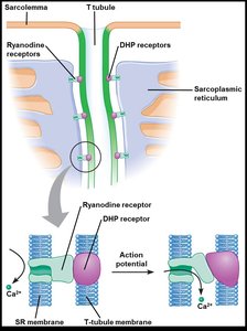
ECC links the electrical signal from the NMJ to muscle contraction. The action potential travels along the sarcolemma and T-tubules, triggering Ca++ release from the SR.
DHP and ryanodine receptors: Mediate Ca++ release from SR.
Ca++ binds troponin: Exposes myosin-binding sites on actin.
Muscle Contraction Types and Mechanics
Motor Units and Muscle Twitch
A motor unit consists of one motor neuron and all the muscle fibers it innervates. A muscle twitch is the response of a muscle to a single stimulus.
All-or-none principle: All fibers in a motor unit contract if the neuron fires.
Phases of twitch: Latent period, contraction phase, relaxation phase.

Types of Muscle Contractions
Muscle contractions can be classified as isotonic (muscle changes length) or isometric (muscle length remains constant).
Isotonic: Concentric (shortening), eccentric (lengthening).
Isometric: Muscle generates force without changing length.
Force Generation and Muscle Performance
Factors Affecting Force Generation
Force generation in muscle depends on crossbridge formation, frequency of stimulation, fiber diameter, and fiber length. Recruitment of more and larger motor units increases force.
Frequency of stimulation: Higher frequency leads to summation and tetanus.
Fiber diameter: Thicker fibers generate more force.
Fiber length: Optimal length maximizes force (length-tension relationship).
Motor unit recruitment: Nervous system activates more units for greater force.
Velocity of Shortening
The speed at which a muscle shortens depends on the load. Heavier loads decrease the velocity and duration of shortening.
Latent period increases with heavier loads.
Velocity decreases as load increases.
Muscle Metabolism and Fiber Types
ATP Production in Muscle
Muscle cells produce ATP through glycolysis, the Krebs cycle, and oxidative phosphorylation. The creatine-phosphate system provides rapid ATP replenishment during intense activity.
Glycolysis: Fast ATP production, low yield.
Oxidative phosphorylation: Slow, high yield.
Creatine kinase reaction:
Types of Skeletal Muscle Fibers
Muscle fibers are classified by their speed of contraction and metabolic properties. Most muscles contain a mix of fiber types.
Slow Oxidative (Type I): High endurance, high oxidative capacity, small diameter.
Fast Oxidative-Glycolytic (Type IIA): Intermediate properties.
Fast Glycolytic (Type IIB): High force, low endurance, large diameter.
Fiber Type | Oxidative Capacity | Glycolytic Capacity | Speed | Fatigue Resistance | Diameter |
|---|---|---|---|---|---|
Slow Oxidative (Type I) | High | Low | Slow | High | Small |
Fast Oxidative-Glycolytic (Type IIA) | Intermediate | Intermediate | Intermediate | Intermediate | Intermediate |
Fast Glycolytic (Type IIB) | Low | High | Fast | Low | Large |
Adaptations to Exercise
Exercise training induces changes in muscle fiber properties. Endurance training enhances oxidative capacity, while resistance training increases muscle hypertrophy. Disuse leads to muscle atrophy.
Endurance exercise: Fast-glycolytic fibers shift toward oxidative properties.
Resistance exercise: Muscle hypertrophy, shift toward glycolytic properties.
Disuse: Muscle atrophy.
